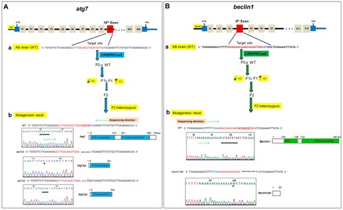
Generation of atg7 and beclin1 zebrafish mutants. (A) The procedure of atg7 targeted mutagenesis. (a) Schematic representation of the zebrafish atg7 target site and generation mating between atg7 F1 heterozygous male (blue circle) and female (red circle) to produce F2 generation. The thin line and grey boxes represent the introns and exons, respectively, and the red box represents the target 10th exon. The sgRNA target sequence is shown in red, followed by a PAM sequence “TGG” shown in blue. (b) Genotyping and illustration of the deduced protein structure of wild-type atg7 and two mutated atg7. The black bar indicates the target sequence of a atg7 wild type (n = 5 bp) in the target site and the black star refers to the deletion part in mutants. (B) The procedure of beclin1 targeted mutagenesis. (a) Schematic representation of the zebrafish beclin1 target site and generation mating between beclin1 F1 heterozygous male (blue circle) and female (red circle) to produce F2 generation. The red box represents the target 4th exon. The sgRNA target sequence is shown in red, followed by a PAM sequence “TGG” shown in blue. (b) Genotyping and illustration of the deduced protein structure of wild-type beclin1 and the mutated beclin1. The black bar indicates the target sequence of a beclin1 wild type (n = 8 bp) in the target site and the black star refers to the deletion part in mutants.

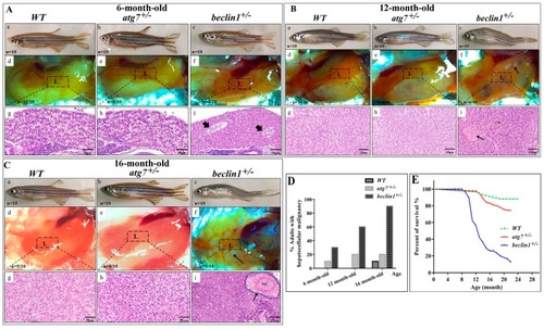
Beclin1 heterozygosity leads to liver disorders in adult males. (A) (ac) Representative pictures of 6-month-old male zebrafish. (df) The liver phenotypes in dissected males. (gi) hematoxylin and eosin (H&E) histological staining of zebrafish livers and the beginning of bile ductus sequestration (arrowheads). (B) (ac) Representative pictures of 12-month-old male zebrafish. beclin1+/− has enlarged the belly and curved the body. (df) The liver phenotypes in dissected males. Hepatic ulcers (black arrow) were observed in the beclin1+/− males. (gi) H&E histological assay showing the tumor cells cords and bile sequestrations (black arrow) in beclin1+/−. (C) (ac) Representative pictures of 16-month-old male zebrafish. (df) The liver phenotypes in dissected males. beclin1+/− liver had a hemorrhagic and necrotic appearance distributed on the inferior surface of the liver (black arrow). (gi) H&E histological assay showing the development of confocal necrosis at 16-month-old male beclin1+/− (black arrow). NC: Necrosis. (D) Prevalence of microscopic hepatocellular malignancy. n = 10 for each experimental group. (E) Meire Kaplan graphs depicting the survival rate of the three reared strains. n = 50 for each experimental group. Beclin1+/− exhibited lower survival rate than WT at 12- and 16-month-old respectively (p < 0.0001). L: liver, the dotted frame represents liver histology in the underlined pictures.

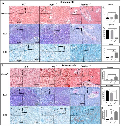
Defects of hepatic energy metabolism in male Beclin1+/− zebrafish. (A) Histological assessment of hepatocytes in 12-month-old male zebrafish. (ad) Representative photomicrographs of Masson’s trichrome staining and the quantification of fibrosis. (eh) Representative photomicrographs of Periodic Acid-Schiff (PAS) staining and glycogen content showing that glycogen packed in the hepatocytes of wild type (WT) and atg7+/− and lacked in beclin1+/− zebrafish; notice the beginning of hepatocellular malignancy (black star) around bile ducts. (il) Oil Red O (ORO) staining and lipid quantification revealing the alternations in fat droplet formation in beclin1+/− hepatocytes. (B) Histological assessment of hepatocytes in 16-month-old male zebrafish. (ad) Representative photomicrographs Masson’s trichrome staining and fibrosis quantification with few fibrils lining the bile duct (arrowhead) and tumor-like cells (black stars) in beclin1+/−. (eh) Representative photomicrographs of PAS staining with sufficient glycogen in WT and atg7+/− and sever glycogen reduction in beclin1+/−, notice the necrotic development around bile ducts (black stars). (il) ORO stain and lipid quantification showing hepatocytes hypertrophy with lipid accumulation and fatty clouds in beclin1+/− zebrafish. Scale magnification is shown in pictures and data are expressed as mean ± SD. * p < 0.05, ** p < 0.01, and *** p < 0.001. CV: Central Vein; BD: Bile Duct; NC: Necrosis.

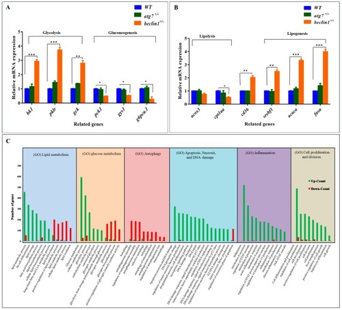
Effects of beclin1 heterozygosity on the hepatic glucose/lipid flux at the genetic level. Expressions of mRNA in 16-month-old zebrafish evaluated by qRT-PCR for glycolysis and gluconeogenesis related genes (A) and lipolysis and lipogenesis related genes (B) indicating the disturbance of glycogen and lipid metabolism. (C) Gene Ontology (GO) analysis of differential expression genes (DEGs) in the liver between beclin1+/− and WT. The y-axis represents the number of genes of the biological processes in the x-axis. The numbers of upregulated DEGs (green bars) and downregulated DEGs (red bars) are shown. Data are expressed as mean ± SD. * p < 0.05, ** p < 0.01, and *** p < 0.001.

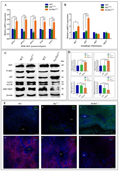
Dysregulated phosphoinositide-3-kinase (PI3K), the serine-threonine protein kinase (AKT) and autophagy pathways in the liver of beclin1+/− zebrafish. Expressions of mRNA in 16-month-old zebrafish evaluated by qRT-PCR for PI3K subunits and anti-apoptotic genes (A) and autophagy-related genes (B). (C) Representative immunoblots showing the relative protein expression of Akt, P-Akt, P62, LC3-I-II, Atg5-Atg12 conjugate. β-actin was used as vehicle control. (D) The ratio of P-Akt/Akt, P62/β-actin, LC3-II/LC3-I, and Atg5-atg12/β-actin quantified by NIH software Image J. (E) (ac) Immunofluorescence of P62 showing protein increasing around the necrosis tissue of beclin1+/−.(df) Immunofluorescence of liver tissues probed against LC3-II revealed a severe reduction in beclin1+/−. CV: central vein; NC: Necrosis. Data expressed as mean ± SD. * p < 0.05, ** p < 0.01, and *** p < 0.001.

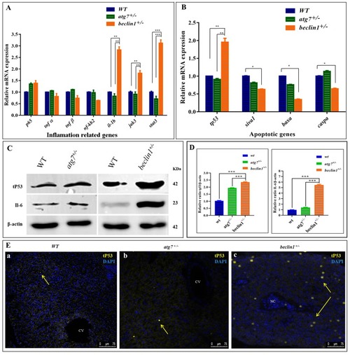
Abnormal apoptosis and inflammation response in beclin1+/− liver. Expressions of mRNA in 16-month-old zebrafish evaluated by qRT-PCR for inflammation-related genes (A) and apoptotic-related genes (B). (C) Representative immunoblots showed the relative protein expression of tP53, IL-6. β-actin was used as the vehicle control. (D) The ratio of tP53/β-actin and IL-6/β-actin evaluated by NIH software Image J. (E) Immunofluorescence of liver tissues probed against mutated tP53. The nuclei of the necrosis hepatocytes indicated the genomic instability and DNA damage in tumor cells (yellow arrows). CV: central vein; NC: Necrosis. Data expressed as mean ± SD. * p < 0.05, ** p < 0.01, and *** p < 0.001.

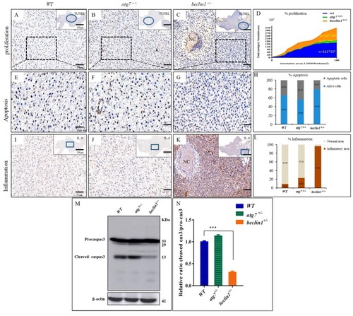
Beclin1 heterozygosity induces cellular proliferation in the hepatic tissue with lower apoptosis and a higher risk of inflammation. (AC) In situ detection of apoptotic cells by TUNEL staining by higher magnification of the blue circles. Brown apoptotic cells around necrosis (black arrow) are shown. (D) The mean cell number per unit area was quantified by NIH Image J. The results were normalized from three independent slides of the same area. (EG) Magnified views of the boxed regions in (AC). (H) Percentage of apoptotic cells according to E-G. The data are shown as the statistical results of three independent experiments, three fields scored per condition. (IK) Immunohistochemistry assay of liver tissues blocked against IL-6 showed in higher magnification of the blue frames. (L) The signals in (IK) were scanned with a Pannoramic MIDI scanner to perform image quantification and analysis. Results were summarized from three independent replicates and data were expressed by mean. (MN) Western blot analysis and quantified protein expression showed that cleaved-caspase3 were downregulated in the beclin1+/− livers in comparison with the WT or atg7+/−. β-actin was used as an internal control. NC: Necrosis. * p < 0.05, ** p < 0.01, and *** < 0.001.

Unillustrated author statements

Acknowledgments
This image is the copyrighted work of the attributed author or publisher, and ZFIN has permission only to display this image to its users. Additional permissions should be obtained from the applicable author or publisher of the image. Full text @ Int. J. Mol. Sci.