- Title
-
A new mib allele with a chromosomal deletion covering foxc1a exhibits anterior somite specification defect.
- Authors
- Hsu, C.H., Lin, J.S., Lai, K.P., Li, J.W., Chan, T.F., You, M.S., Tse, W.K., Jiang, Y.J.
- Source
- Full text @ Sci. Rep.
|
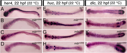
The new alleles show similar Notch molecular phenotypes to those of previous mib mutants.All embryos were raised at 22 °C and flat-mounted WISH performed at 22 hpf. A-D are flat-mounted her4 WISH. (A) WT embryos expressed her4 in brain and neural tube; (B) mibnn2000, (C) mibnn2001 and (D) mibnn2002 mutants all showed a down-regulation of her4 expression. E-H are flat-mounted huc WISH. (E) WT embryos expressed huc in cells of brain and neural tube; (F) mibnn2000, (G) mibnn2001 and (H) mibnn2002 mutants all showed an up-regulation of huc expression. I-L are flat-mounted dlc WISH. (I) WT embryos expressed dlc in PSM, formed somites and cell clusters in the hindbrain; (J) mibnn2000, (K) mibnn2001 and (L) mibnn2002 mutants all showed a salt-and-pepper dlc pattern in the nascent somites and anterior PSM. Note that the dlc expression in formed somites was lost in (L) mibnn2002 mutants. EXPRESSION / LABELING:
PHENOTYPE:
|
|
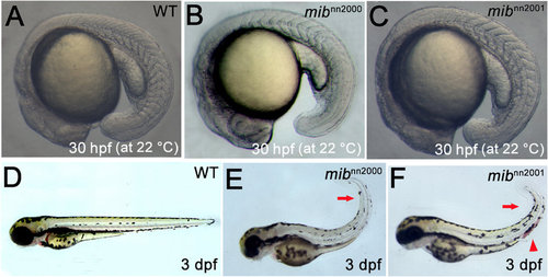
Morphological phenotypes of mibnn2000 and mibnn2001 mutants. A-C are the lateral views of 30 hpf embryos raised at 22 °C. While (A) WT embryos showed about 20 somites at 30 hpf, (B) mibnn2000 homozygotes showed about 11 recognizable somites and (C) mibnn2001 homozygotes showed about 14 somites. D-F are lateral views of 3 dpf embryos at 28.5 °C. Compared to (D) WT embryos, (E) mibnn2000 and (F) mibnn2001 mutants showed a curly tail (arrows), hemorrhage (arrowhead) and a reduction of tail pigmentation (more prominent in E). PHENOTYPE:
|
|
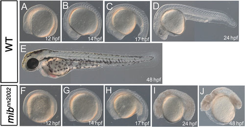
mibnn2002 mutants show phenotypes distinct from those of typical mib mutants. A-E are lateral views of WT embryos. F-J are lateral views of mibnn2002 homozygotes. A and F are at 12 hpf; B and G are at 14 hpf; C and H are at 17 hpf; D and I are at 24 hpf; E and J are at 48 hpf. No visible somites can be observed from the lateral view of mibnn2002 homozygotes at (F) 12 hpf and (G) 14 hpf. Visible somite-like structures can be discerned from the lateral view of mibnn2002 homozygotes at (H) 17 hpf and (I) 24 hpf. Cell death can be spotted in mibnn2002 homozygotes at (I) 24 hpf and (J) 48 hpf. Edema was obvious in mibnn2002 homozygotes at (J) 48 hpf. PHENOTYPE:
|
|
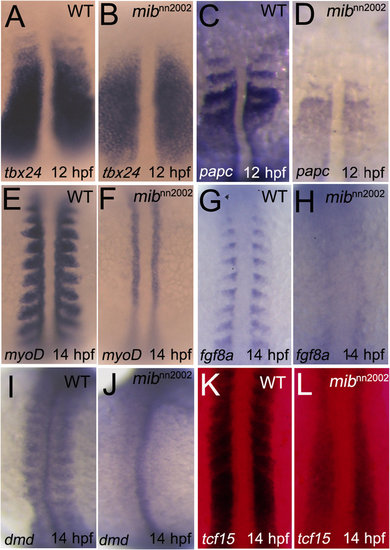
Somite/boundary markers are lost in mibnn2002 mutants. (A) WT embryos and (B) mibnn2002 mutants were stained with tbx24 at 12 hpf. (C) WT embryos and (D) mibnn2002 mutants were stained with papc at 12 hpf. (E) WT embryos and (F) mibnn2002 mutants were stained with myoD at 14 hpf. (G) WT embryos and (H) mibnn2002 mutants were stained with fgf8a at 14 hpf. (I) WT embryos and (J) mibnn2002 mutants were stained with dystrophin (dmd) at 14 hpf. (K) WT embryos and (L) mibnn2002 mutants were stained with tcf15 at 14 hpf, where DsRed filter was used to improve the signal to noise ratio of NBT/BCIP staining under transmitted light. All embryos were mounted in dorsal view with head to the top. tbx24 and papc were expressed in PSM, though the anterior segmental pattern was not observed in the region of nascent somites in mibnn2002 mutants. The expression of myoD, fgf8a, and dystrophin were not detected in the region where somites normally formed. tcf15 was weakly expressed in mibnn2002 mutants and no segmental pattern can be discerned. EXPRESSION / LABELING:
PHENOTYPE:
|
|
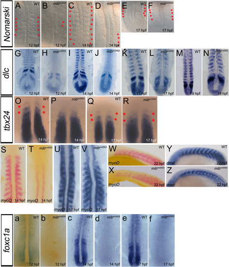
Somite segmentation and specification recover without foxc1a expression. A-F are dorsal views of WT embryos and mibnn2002 mutants. Small round-shaped structures can be observed in (B) mibnn2002 homozygotes at 12 hpf. The newly-generated somite-like structures increased when mibnn2002 homozygotes grew to (D) 14 hpf and (F) 17 hpf. G-N are flat mounts of WT embryos and mibnn2002 mutants stained with dlc. (H) The expression of dlc in the region where somites normally formed was not observed in mibnn2002 homozygotes at 12 hpf. (J, L and N) dlc progressively appeared after 14 hpf in the newly-generated somites. O-R are dorsal views of WT embryos and mibnn2002 mutants stained with tbx24. The segmental tbx24 (marked with red asterisks) appeared progressively in mibnn2002 homozygotes. S-V are flat mounts of WT embryos and mibnn2002 mutants stained with myoD. W and X are lateral views of WT embryos and mibnn2002 mutants stained with myoD. The expression of myoD in formed somites started to emerge after 14 hpf as what was observed in dlc. As embryos developed, (V) the expression of myoD progressively appeared in the region where somites normally formed, except the anterior segments. (X) The cells that expressed myoD still split into dorsal and ventral myotomes even where somite boundaries were not formed. Y and Z are lateral views of WT embryos and mibnn2002 mutants stained with dystrophin (dmd) at 22 hpf. The expression of dystrophin can be observed in formed somites. Note that the somite gap delineated by dystrophin in formed somite region is similar between (Y) WT embryos and (Z) mibnn2002 mutants. a-f are dorsal views of WT embryos and mibnn2002 mutants stained with foxc1a. The expression of foxc1a in mibnn2002 homozygotes are not detected in all the stages examined (b 12 hpf; d 14 hpf and f 17 hpf). EXPRESSION / LABELING:
PHENOTYPE:
|
|
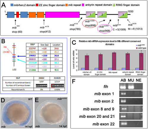
mib gene is deleted in mibnn2002 mutants. (A) Diagram of the mutations of different mib alleles. The translation of mib in mibnn2000 and mibnn2001 mutants stops at amino acids 5′ to ring finger domain 3 (RF3). (B) The linkage analysis result of mibnn2002 mutants. The upper part illustrates the locations of mib gene and the SSLP markers. The lower part demonstrates a close linkage of Z4662 and Z13620 to mib, with a recombination frequency of 0/97 and 2/214, respectively. (C) Real-time PCR results of mib expression in mibnn2002 homozygotes and WT embryos. The blue bars represent the expression level of mib in mibnn2002 homozygotes. The purple bars represent the expression level of mib in WT embryos. There is nearly no mib transcript detected in the mibnn2002 homozygotes. (D) WT embryos and (E) mibnn2002 mutants were stained with mib at 14 hpf, respectively. There is no mib mRNA detected in (E) mibnn2002 homozygous. (F) The results of mib genomic PCR were shown with fih (hif1an) as a positive control. AB represents AB WT samples; MU represents mibnn2002 homozygous samples; NE represents negative controls, where distilled water was used instead of genomic extracts as templates. The results showed that no mib genomic fragments are amplified. EXPRESSION / LABELING:
PHENOTYPE:
|
|
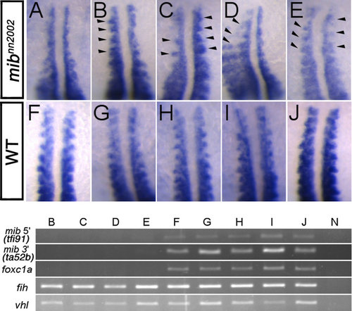
Anterior myoD expression in somites is partially rescued by foxc1a overexpression at 16 hpf.Representative WISH results of (A) non-injected control mibnn2002 mutants, (B-E) 20 pg foxc1a mRNA injected mibnn2002 mutants and (F-J) 20 pg foxc1a mRNA injected WT (siblings) are shown. The rescued somites are marked by arrowheads. myoD expression in the anterior somites of E was rescued symmetrically, while those of B-D were rescued asymmetrically in terms of myoD expression pattern, which may be caused by the unequal segregation of injected foxc1a mRNA. Lower panel shows the genotyping results of the embryos shown in B-J. The genomic fragments of mib 5′ (amplified by tfi91 primer set), mib 3′ (amplified by ta52b primer set) and foxc1a were not detected in B-E, while they were amplified in F-J. fih and vhl were amplified in all samples except for the negative control N. None of the genomic fragments was detected in the negative control. EXPRESSION / LABELING:
PHENOTYPE:
|
|
Cold-sensitive dlcmibnn2002 mutants. Embryos were raised at 22 °C before the desired stage (siblings are with 16 somites or at 27 hpf). Typical salt-and-pepper pattern shown in previous Notch-related mutants was observed in mibnn2002 mutants. Although dlc was still cyclically expressed in the PSM of mibnn2002 mutants, the sharp-striped pattern of dlc |
|
Somite phenotypes of homozygotes (mibta52b/mibta52b) and transheterozygotes (mibta52b/ mibnn2002). Embryos are in lateral view with head to left. While 22 visible somites were observed in the WT embryos (left panel) at 22 ss, only 3 and 7 visible somites were observed in mibta52b homozygotes (middle panel) and transheterozygotes (mibta52b♂ x mibnn2002♀, right panel), respectively. PHENOTYPE:
|
|
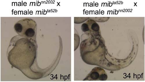
The maternal effect on phenotypes of transheterozygotes (mibta52b/mibnn2002) at 34 hpf. The transheterozygotes have typical mib phenotype at 34 hpf, including less pigmentation and curly tail. Note that the transheterozygous embryos from incrosses of male mibnn2002 carrier and female mibta52b carrier (left) showed a more severe pigmentation phenotype (typical white tail phenotype of ta52b allele17) than those from incrosses of male mibta52b carrier and female mibnn2002 carrier (right). PHENOTYPE:
|
|
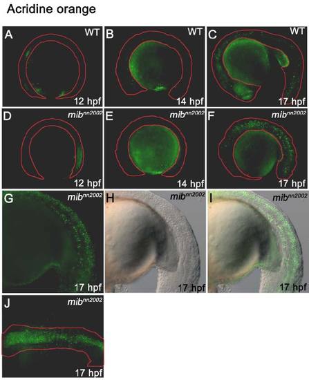
Cell death in mibnn2002 mutants. All the embryos are treated with acridine orange. A-C are wt embryos; D-J are mibnn2002 homozygotes. A and D are at 12 hpf; B and E are at 14 hpf; C and F-J are at 17 hpf. A-I are lateral views with head toward left; J is dorsal view with head toward left. The contours of embryos are outlined by the red lines. (F) The amount of cell death is highly increased in 17 hpf mibnn2002 homozygotes. Cell death was mainly located around (G-I) neural tube and notochord. PHENOTYPE:
|
|
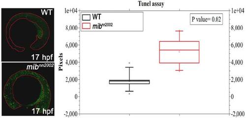
Apoptosis is highly increased in mibnn2002 mutants. TUNEL assay was employed to study the apoptosis in wt embryos and mibnn2002 homozygotes. The lateral view of embryos is shown with head to the left. Apoptosis is increased in mibnn2002 homozygotes at 17 hpf. Analyzed and calculated by Image J, the fluorescent signal increases by 2~3-fold in mibnn2002 homozygous embryos. PHENOTYPE:
|
|
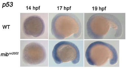
p53 is highly up-regulated in mibnn2002 mutants. p53 WISH was performed in the wt embryos and mibnn2002 homozygotes. Embryos are shown in lateral views with head to the left. The staining of p53 was highly up-regulated in mibnn2002 mutants compared to wt embryos. EXPRESSION / LABELING:
PHENOTYPE:
|
|
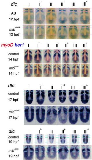
The expression of dlc in mibnn2002 mutants during the stages of 12 hpf, 14 hpf, 17 hpf and 19 hpf at 28.5 °C. dlc WISH was performed in the wt embryos and mibnn2002 homozygotes. Embryos are flat-mounted with head to the top. The cyclically dlc expression was detected in both wt embryos and mutants. No salt-and-pepper pattern was observed. |
|
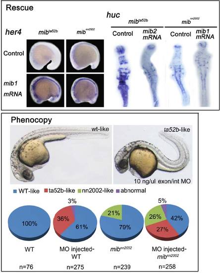
The rescue and phenocopy of mibnn2002 phenotypes. In rescue experiments, her4 WISH was carried out in the mib mRNA (1 ng/µl) injected mibta52b and mibnn2002 embryos. 19 hpf embryos in lateral views with head toward left were shown. The expression of her4 was rescued (increased) by mib mRNA in mibta52b and mibnn2002 homozygotes. huc WISH was carried out in the mib2 mRNA (1 ng/µl) injected mibta52b and mib mRNA (1 ng/µl) injected mibnn2002 embryos. 19 hpf embryos in flat mount with head toward top were shown. huc was rescued (decreased) by mib2 mRNA in mibta52b and mib mRNA in mibnn2002 homozygotes. In phenocopy experiments, mib morpholino injected-wt and -mibnn2002 embryos were examined. Morpholino can successfully convert 36% of wt and 27% mibnn2002 siblings into embryos that exhibit typical mib phenotypes (ta52blike). However, the morpholino failed to convert the mibnn2002 homozygous embryos into embryos having typical mib phenotypes. The ratio of embryos carried mibnn2002 phenotype is 21% without injection and 26% with injection. The ratio of embryos that exhibited mibnn2002 phenotypes was not reduced with the injection of mib morpholino. |

