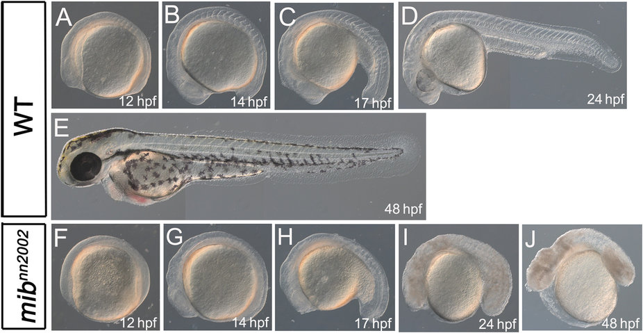
Fig. 3
mibnn2002 mutants show phenotypes distinct from those of typical mib mutants. A-E are lateral views of WT embryos. F-J are lateral views of mibnn2002 homozygotes. A and F are at 12 hpf; B and G are at 14 hpf; C and H are at 17 hpf; D and I are at 24 hpf; E and J are at 48 hpf. No visible somites can be observed from the lateral view of mibnn2002 homozygotes at (F) 12 hpf and (G) 14 hpf. Visible somite-like structures can be discerned from the lateral view of mibnn2002 homozygotes at (H) 17 hpf and (I) 24 hpf. Cell death can be spotted in mibnn2002 homozygotes at (I) 24 hpf and (J) 48 hpf. Edema was obvious in mibnn2002 homozygotes at (J) 48 hpf.

