- Title
-
Notch signaling coordinates cellular proliferation with differentiation during zebrafish fin regeneration
- Authors
- Grotek, B., Wehner, D., and Weidinger, G.
- Source
- Full text @ Development
|
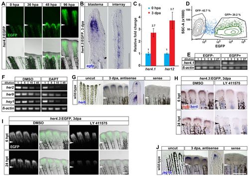
Notch signaling is activated in the blastema during regenerative outgrowth. (A) EGFP fluorescence in her4.3:EGFP transgenics. By 96 hpa, EGFP signal is strongly intensified (exposure times: 0, 36 and 48 hpa for 7 seconds; 96 hpa for 0.5 seconds). (B) Longitudinal sections of her4.3:EGFP regenerates at 3 dpa stained for egfp. There is expression in the proximal but not the distal blastema, and weak expression in interray tissue. (C) her4.1 and her12 levels in 3 dpa regenerates measured by qRT-PCR, shown relative to the level in the distal-most stump segment at 50% fin length (0 hpa). (D) FACS scatterplot showing cell fractions of EGFP+ and EGFP- cells from dissociated 4 dpa her4.3:EGFP regenerates. (E) Endogenous her4.1 transcripts can be detected only by semi-quantitative PCR in the EGFP+ fraction of her4.3:EGFP regenerates. (F) her2, her9 and hey1 expression is downregulated in 3 dpa regenerates treated with DAPT for 6 hours. (G) her6 is expressed in the blastema at 3 dpa. (H) her4.3-driven egfp transcripts (red) and endogenous her6 (blue) are downregulated in regenerates treated with LY411575 6 hours after the start of treatment (hpt) (n=6/6 fins) and not detectable at 24 hpt (n=5/6). (I) her4.3-driven EGFP fluorescence is downregulated in 3 dpa regenerates treated with LY411575 for 24 hours, but not for 6 hours. (J) jag1b is expressed in the blastema at 3 dpa. (A-J) Arrowheads indicate amputation plane. Scale bars: whole mounts, 200 μm; sections, 100 μm. EXPRESSION / LABELING:
|
|
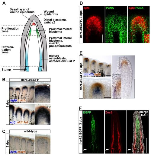
Notch pathway activity is confined to the proliferative zone of the proximal medial blastema. (A) A longitudinal section of a regenerating fin during the outgrowth phase of regeneration (after 48 hpa) showing relevant anatomical structures and expression domains. (B) The her4.3:EGFP reporter (blue) is expressed proximally to msxb and aldh1a2 (red) at 3 dpa. (C) her6 (blue) is expressed proximal to msxb (red). (D) her4.3:EGFP is active in PCNA-positive cells. Confocal images of whole-mount regenerates stained for egfp transcripts and PCNA protein. (E) her4.3:EGFP (red) expression extends further distally than runx2b (blue), and is confined to the medial, runx2b-negative blastema. (F) Confocal images of longitudinal sections of her4.3:EGFP transgenic regenerates stained with Zns5 antibody (labeling all osteoblasts) and anti-GFP show no overlap. (B-E) Arrowheads indicate amputation plane. Scale bars: whole mounts, 200 μm; sections, 100 μm. |
|
Notch signaling is necessary for fin regeneration. (A) Schematic timeline of fin regeneration and experimental treatments. (B) Fins treated with LY411575 starting from the time of amputation fail to regenerate. (C) Regenerate length of fins treated with DMSO or LY411575 from the time of amputation, mean±s.e.m., n=6 fish, 18 fin rays each group (LY at day 6: n=5 fish, 15 fin rays). At all timepoints, except 1 and 2 dpa, regenerate lengths are significantly different (P<0.001) in control and LY411575-treated fins, Student’s t-test. (D) LY411575 treatment from the time of amputation does not affect downregulation of EGFP expression (arrowhead) in the distalmost stump segment of osteocalcin:EGFP transgenic fins. Dashed line indicates amputation plane. (E,F) EGFP pixel intensity plots of representative osteocalcin:EGFP transgenic fin rays at the indicated time points after amputation treated with DMSO (E) or LY411575 (F), showing a decrease in intensity in a region 250 μm proximal to the amputation plane (0 on x-axis, right) and a shift of intensity towards the amputation plane (arrow). (G) Treatment with LY411575 for 4 days starting from 2 dpa is sufficient to interfere with regenerative outgrowth. (H) Regenerate length of fins treated as those in G at 2 dpa (prior to the start of treatment) and at 6 dpa, 4 days after the start of treatment (dpt). Mean±s.e.m. n=6 fins, 18 fin rays. ***P<0.001. (I) Confocal images of anti-BrdU immunofluorescence in the mesenchymal compartment of fin rays at 3 dpa treated with DMSO or LY411575 for 6 hours. (J) Number of BrdU-positive cells in the distal 200 μm of the mesenchyme of fins treated as in I. Mean±s.e.m. DMSO: n=7 fish, 27 blastemas; LY 411575: n=7 fish, 26 blastemas. ***P<0.001. (K) The blastema markers msxb (6/6 fins) and ilf2 (5/6 fins) are downregulated in regenerates treated with LY411575 for 24 hours starting at 2 dpa. (B-K) Red arrowheads indicate amputation plane. Scale bars: 200 μm in B,D,G,K; 100 μm in I. EXPRESSION / LABELING:
|
|
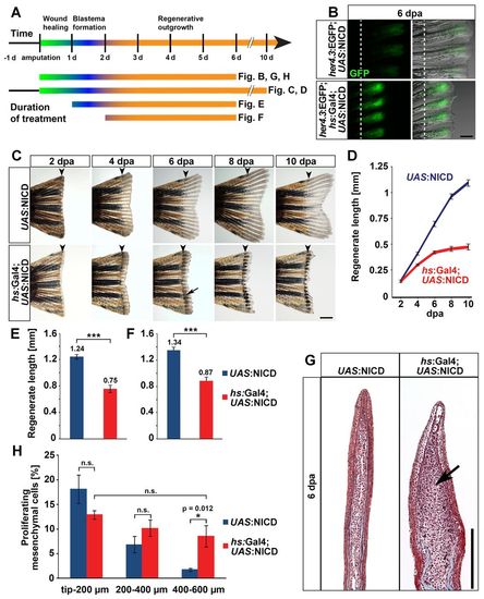
NICD overexpression expands the proliferative blastema, yet stalls fin regeneration. (A) Schematic timeline of fin regeneration and experimental treatments. (B) Enhancement of EGFP expression in the blastema of her4.3:EGFP; hs:Gal4; UAS:NICD triple transgenic fins at 6 dpa after repeated heatshocks starting at the time of amputation. Dashed line indicates amputation plane. Scale bar: 200 μm. (C) Inhibition of regeneration in hs:Gal4; UAS:NICD fish heatshocked repeatedly starting 1 day prior to fin amputation. Pigment cells cluster in the stalled regenerate (arrow). Arrowheads indicate amputation plane. Scale bar: 500 μm. (D) Regenerate lengths of fins treated as in C; mean±s.e.m. Length differences at all timepoints, except 2 dpa, are highly significant (P<0.001). UAS:NICD: n=8 fins, 24 fin rays; hs:Gal4; UAS:NICD: n=12 fins, 36 fin rays. (E,F) NICD overexpression starting at 1 dpa (E) or 2 dpa (F) is able to delay fin regeneration, mean±s.e.m. n=8 fins, 24 fin rays each group. ***P<0.001. (G) Masson’s trichrome staining reveals expansion of the mesenchymal compartment of regenerates (arrow) after sustained NICD overexpression for 6 days. Scale bar: 200 μm. (H) Percentage of BrdU-positive mesenchymal cells in three 200 μm regions along the distal-proximal axis of UAS:NICD and hs:Gal4; UAS:NICD fins heatshocked repeatedly for 6 days. NICD overexpression increases proliferation in the scarcely proliferating proximal 400-600 μm domain (‘differentiation zone’). n=5 fish, six sections. *P<0.05. EXPRESSION / LABELING:
|
|
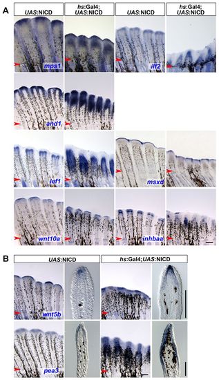
NICD overexpression causes expansion of proximal mesenchymal and epidermal compartments. (A,B) Whole-mount in situ hybridization of hs:Gal4; UAS:NICD and UAS:NICD fins heatshocked repeatedly from 0 hpa to 6 dpa with the indicated markers reveals massive proximal expansion of proximal, but not distal, blastemal and epidermal domains upon NICD overexpression. mps1=6/6 fins; ilf2=6/6; and1=9/11; lef1=7/7; wnt10a and inhbaa=4/5; wnt5b and pea3=5/6. Arrowheads indicate the amputation plane. Scale bars: whole mounts, 200 μm; sections, 100 μm. EXPRESSION / LABELING:
|
|
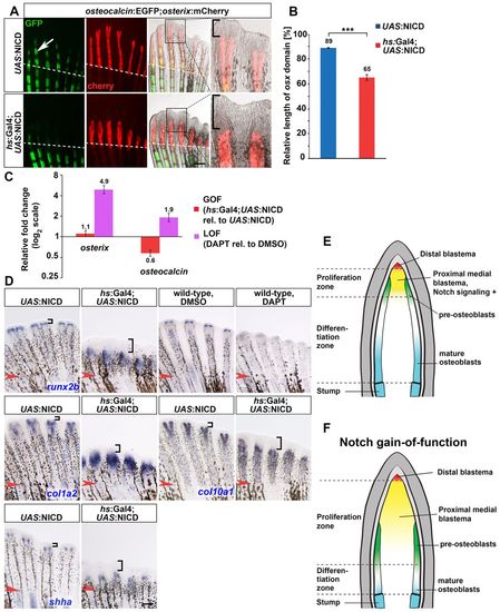
Notch signaling negatively regulates osteoblast differentiation in the regenerating fin. (A) Altered bone marker expression in the regenerate of osteocalcin:EGFP; osterix:mCherry; hs:Gal4; UAS:NICD quadruple transgenic fish heatshocked repeatedly for 6 days. Dashed line indicates amputation plane. Scale bar: 200 μm. Arrows and brackets indicate osteocalcin: EGFP. (B) The fraction of the regenerate expressing osterix:mCherry in fish treated as in A. Mean±s.e.m. n=7 fins, 31 fin rays. ***P<0.001. (C) qRT-PCR shows upregulation of osteocalcin and osterix at 6 dpa in regenerates treated with DAPT for 4 days from 2 dpa onwards (loss of function), whereas osteocalcin is downregulated upon sustained NICD overexpression (gain of function) for 6 days. Mean±s.d., n=15 regenerates transgenics, 10 drug treatments. (D) Whole-mount in situ hybridization of the indicated markers in 6 dpa regenerates overexpressing NICD or treated with DAPT for 6 days. n=5/6, runx2b (hs:Gal4; UAS:NICD); n=12/13, runx2b (DAPT); n=6/6, col1a2; n=4/5, col10a1; n=5/7, shha. Arrowheads indicate amputation plane. Brackets indicate distal region devoid of marker expression. (E,F) Model depicting the spatial distribution of Notch signaling-positive cells in a longitudinal section of a wild-type fin regenerate (E) and the patterning consequences of Notch gain of function (F). EXPRESSION / LABELING:
|
|
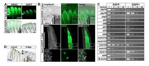
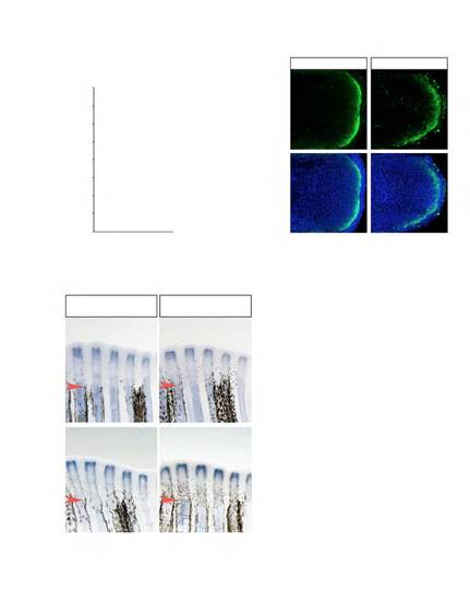
Expression of a transgenic reporter for Notch signaling and Notch receptors and ligands during fin regeneration. (A) In her4.3:EGFP transgenic regenerates treated with DAPT for 24 hours (24 hpt), EGFP fluorescence is reduced relative to DMSOtreated fins (n=3/3). (B) EGFP fluorescence in her4.3:EGFP transgenic regenerates is detectable in a few scattered cells in the wound epidermis. Top panel: whole-mount image of regenerates at 2 dpa. Bottom panel: confocal images of longitudinal sections of regenerates stained with GFP antibody and DAPI at 3 dpa. (C) Semi-quantitative PCR of the indicated genes on serial dilutions of cDNA derived from the EGFP-positive and EGFP-negative cellular pools of FACS-sorted her4.3:GFP regenerates at 3 dpa. (D) lunatic fringe (lfng) expression, as detected by whole-mount in situ hybridization, is upregulated in the regenerating fin at 3 dpa relative to uncut fins. Arrowheads indicate amputation plane. Scale bars: 200 μm. |
|
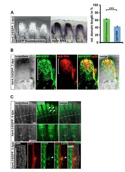
Difference in transcript and protein expression pattern of the her4.3:EGFP reporter. (A) Length of the domain positive for EGFP fluorescence along the proximodistal axis is significantly larger than that of the domain positive for egfp RNA, as detected by whole-mount in situ staining in the same fins rays of her4.3:EGFP transgenics. In the right panel, the domain lengths relative to the length of the entire regenerate are plotted (2nd, 3rd and 4th lateral ray of each lobe was measured, n=5 regenerates, ***P<0.001 t-test). (B) EGFP protein-positive cells extend further proximally than egfp RNA-positive cells in her4.3:EGFP regenerates at 3 dpa. Confocal images of the fluorescent signal of Fast Red precipitates in whole-mount regenerates stained for egfp transcripts and of anti-GFP immunofluorescence. Scale bar: 100 μm. (C) EGFP fluorescence in her4.3:EGFP transgenic regenerates is detectable in the forming joint regions (arrowheads). Top panel: whole-mount image of 4 dpa regenerate. Bottom panel: confocal image of longitudinal sections of 3 dpa her4.3:GFP transgenic regenerates stained with anti-GFP antibody, anti-Zns5 antibody (labeling all osteoblasts) and DAPI. |
|
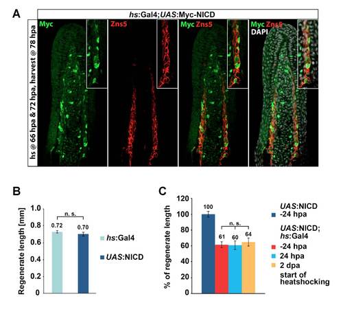
NICD overexpression using the hs:Gal4; UAS:NICD system. (A) Mosaic Myc-NICD expression in regenerates of hs:Gal4; UAS:NICD double transgenics at 78 hpa following 2 heat shocks at 66 hpa and 72 hpa. Confocal image of longitudinal sections stained with anti-Myc antibody, anti-Zns5 antibody (labeling all osteoblasts) and DAPI. (B) Heatshock-induced Gal4 overexpression from amputation until 6 dpa in hs:Gal4 transgenic fish has on its own no influence on regenerate length when compared with fish carrying the UAS:NICD transgene only, mean±s.e.m. n=11 fins, 33 fin rays each group. n.s., not significant, Student’s t-test. (C) NICD overexpression in hs:Gal4; UAS:NICD double transgenic fish inhibits fin regeneration to a similar extent irrespective of the onset of NICD overexpression, i.e. 24 hours before amputation, 24 ours after or 2 days after amputation, as revealed by quantification of the percentage of regrown fin tissue at 6 dpa. n.s., not significant, Student’s t-test. |
|
Short-term NICD overexpression has no effect on blastema proliferation or on fin patterning. (A) NICD overexpression in hs:Gal4; UAS:NICD double-transgenic fish for 24 hours starting at 3 dpa does not induce cell proliferation, as revealed by BrdUincorporation. n= 6 fish (control), 22 blastemas; n= 8 fish (gain of function), 29 blastemas, n.s., not significant, Student’s t-test. (B) NICD overexpression in hs:Gal4; UAS:NICD double transgenic fish for 6 days starting immediately after amputation does not expand the distal-most blastema marked by anti-Aldh1a immunofluorescence detected in confocal images. n= 6/6 (control), n=5/6 (gain of function). (C) NICD overexpression in hs:Gal4; UAS:NICD double transgenic fish for 12 hours starting at 3 dpa does not alter lef1 or runx2b expression. n=7 fins (all groups). |

