FIGURE SUMMARY
Title

Nexmifa Regulates Axon Morphogenesis in Motor Neurons in Zebrafish

Authors
Zheng, Y.Q., Suo, G.H., Liu, D., Li, H.Y., Wu, Y.J., Ni, H.
Source
Full text @ Front. Mol. Neurosci.

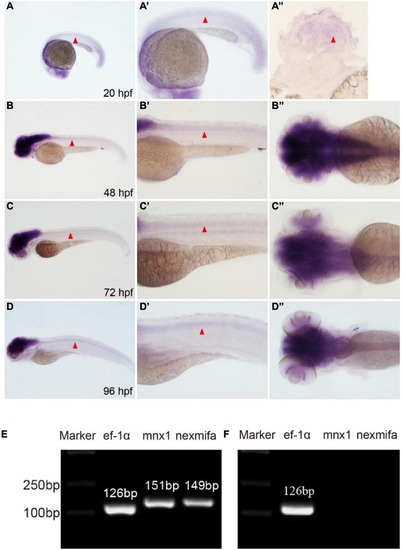
Nexmifa expression analysis in spinal cord and motor neurons. (A) At 20 hpf, in situ hybridization nexmifa signals are localized in brain and spinal cord (lateral view). (A’) Partially magnified image of (A). (A”) The trunk transverse section of the embryo. (B) At 48 hpf, in situ hybridization nexmifa signals are localized in the brain and spinal cord (lateral view). (B’) Partially magnified image of (B). (B”) Dorsal view of the brain. (C) At 72 hpf, in situ hybridization nexmifa signals are localized in the brain and spinal cord (lateral view). (C’) Partially magnified image of (C). (C”) Dorsal view of the brain. (D) At 96 hpf, in situ hybridization nexmifa signals are localized in the brain and spinal cord (lateral view). (D’) Partially magnified image of (D). (D”) Dorsal view of the brain. (E) RT-PCR results on mnx1-GFP sorted cells. Nexmifa is expressed in selected neuron cells from the Tg (mnx1: EGFP) line. (F) The result of the RT-PCR on Kdrl-EGFP sorted cells. No signals of nexmifa and mnx1 are detected in the GFP-positive cells sorted from the Tg(kdrl:EGFP) line.

EXPRESSION / LABELING:
Genes:
Fish:
Anatomical Terms:
Stage Range: 20-25 somites to Day 4

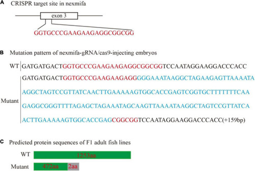
Generation of the zebrafish nexmifa mutant using the CRISPR/Cas9 system. (A) Schematic showing the targeting site of the sgRNA in the third exon of nexmifa. (B) Mutation pattern of nexmifa-gRNA/cas9-injected embryos. Red letters represent the sgRNA sequence. Blue letters represent the additional 159 bp nucleotide sequence. (C) Schematic showing the predicted protein encoded by the mutated allele. Frameshift mutations resulted in truncated proteins. The gray rectangle indicates the wrong coded amino acid sequences.

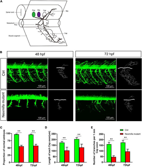
Nexmifa affects motor neuron morphogenesis in nexmifa mutant zebrafish embryos. (A) The schematic for three different primary motoneurons (CaP, MiP, and RoP) in fish. (B) Confocal imaging of primary motor neurons in control and nexmifa mutant groups at 48 and 72 hpf. Scale bar = 100 μm (C) The percentage of embryos with normal PMNs in control and nexmifa mutants at 48 hpf (n = 125 and 240, respectively) and 72 hpf (n = 132 and 235, respectively). (D) Cap length in control and nexmifa mutants at 48 hpf (n = 20 and 31, respectively) and 72 hpf (n = 15 and 28, respectively). (E) Number of branches per 1 mm Cap axon in control and nexmifa mutants at 48 hpf (n = 8 and 10, respectively) and 72 hpf (n = 9 and 12, respectively). Bar represent the mean ± standard deviation (SD). **p < 0.01.

Motor defects in nexmifa mutant zebrafish embryos at 7 dpf. (A) Thirty minutes of free movement trajectories of control and nexmifa mutants. (B) Quantification of swimming distances of control and nexmifa mutants at 7 dpf per 5 min (n = 22 and 24, respectively). Each point represents the mean ± standard error of the mean (SEM). *p < 0.05 and **p < 0.01.

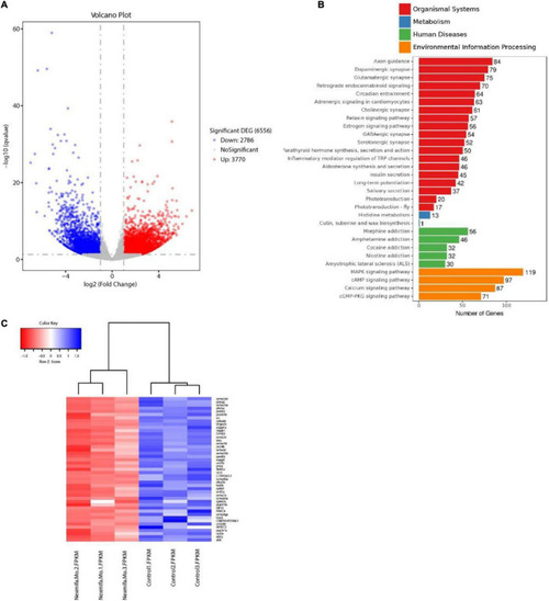
Transcriptomics profiling in nexmifa morphant and wild-type zebrafish. (A) Volcano map showing DEGs in control and nexmifa morphants. Red and blue spots represent up-regulated and down-regulated genes, respectively. (B) KEGG pathway annotation of DEGs in control and nexmifa morphants. (C) Heatmap pathway differences between control and nexmifa morphants showing down-regulated genes in axon guidance.

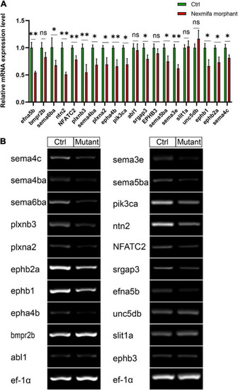
The expression of 20 down-regulated DEGs. (A) Expression of 20 down-regulated DEGs between Ctrl and nexmifa morphant by qRT-PCR. Bars represent the mean ± standard deviation (SD). *p < 0.05 and **p < 0.01. ns: non-significant. (B) Expression of 20 down-regulated DEGs between Ctrl and nexmifa mutant by RT-PCR.

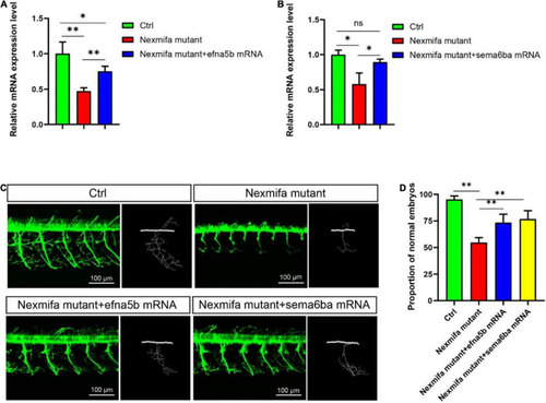
Efna5b and sema6ba overexpression rescues motor neuron defects in nexmifa morphant embryos. (A) Expression of efna5b after injection of efna5b mRNA in nexmifa mutant embryos at 72 hpf. (B) Expression of sema6ba after injection of sema6ba mRNA in nexmifa mutant embryos at 72 hpf. (C) Abnormal PMNs in nexmifa morphant zebrafish were rescued by injecting efna5b or sema6ba mRNA. (D) Percentage of zebrafish embryos with normal Cap primary motor neurons (n = 150, 242, 250, and 257, respectively). *p < 0.05 and **p < 0.01. ns: non-significant.

Efna5b and sema6ba overexpression rescues the impaired motility in nexmifa mutant embryos at 7 dpf. (A) Thirty minutes of free movement trajectories among control, nexmifa mutants, nexmifa mutant + efna5b mRNA and nexmifa mutant + sema6ba mRNA. (B) Quantification of swimming distances among control, nexmifa mutants, nexmifa mutant + efna5b mRNA and nexmifa mutant + sema6ba mRNA at 7 dpf per 5 min (n = 20, 24, and 24, respectively). Each point represents the mean ± standard error of the mean (SEM). **p < 0.01.

Acknowledgments
This image is the copyrighted work of the attributed author or publisher, and ZFIN has permission only to display this image to its users. Additional permissions should be obtained from the applicable author or publisher of the image. Full text @ Front. Mol. Neurosci.