- Title
-
Overexpression of SOX4 correlates with poor prognosis of acute myeloid leukemia and is leukemogenic in zebrafish
- Authors
- Lu, J.W., Hsieh, M.S., Hou, H.A., Chen, C.Y., Tien, H.F., Lin, L.I.
- Source
- Full text @ Blood Cancer J
|
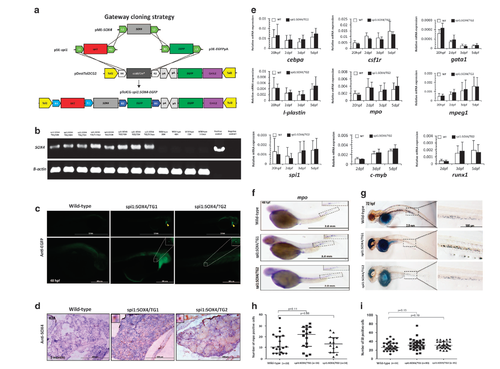
Generation and characterization of the Tg(spi1:SOX4-EGFP) transgenic zebrafish. (a) Schematic diagram showing the LR recombination reaction used to generate the expression constructs, including three entry clones (p5E-spi1, pME-SOX4 and p3E-EGFPpA) and a destination vector (pDestTol2CG2) that contains the cmlc2:EGFP-pA expression cassette. The final construct (pTolCG-spi1:SOX4-EGFP) is shown at the bottom of the figure. (b) Results of semiquantitative PCR showing the expression of SOX4 in TGs and wild-type embryos at 20, 48 and 72 h post fertilization (HPF) and at 5 days. Negative control: non-template; Positive control: pTolCG-spi1:SOX4-EGFP plasmid. (c) Whole-mount immunohistochemical staining (IHC) showed EGFP-positive cells in the heart (yellow arrow) as well as around the yolk sac and caudal hematopoietic tissue (CHT) at 48 HPF. The panel shows GFP+ cells at a higher magnification. (d) Tissue sections showing SOX4-positive cells in the KM of Tg(spi1:SOX4-EGFP) fish but not in those of wild-type fish. The panel shows SOX4-positive cells at a higher magnification. (e) Results from quantitative reverse transcription-PCR (RT-PCR) analysis of hematopoietic marker genes in SOX4 transgenic zebrafish embryos and in wild-type fish embryos. Data are presented as the mean±s.e.m. from three independent experiments. (f, g) Results from whole-mount in situ hybridization (WISH) of mpo and Sudan black (SB) staining showing mpo-positive cells in CHT at 48 HPF and SB-positive cells in CHT at 72 HPF, respectively (magnification: × 40). The panel shows positive cells at a higher magnification (× 100), respectively. Quantification of mpo-positive cells (h) and SB-positive cells (i). Differences among variables were assessed using Student’s t-test. EXPRESSION / LABELING:
PHENOTYPE:
|
|
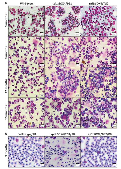
Morphological analysis of various blood cell types harvested from the KM and peripheral blood of Tg(spi1:SOX4-EGFP) zebrafish at indicated age. Tg(spi1:SOX4-EGFP) KM smears showed myeloid hyperplasia with increased production of myeloblasts in an age-dependent manner (a) and their peripheral blood smears showed immature myeloid cells (b). Conversely, wild-type fish showed normal hematopoiesis with adequate maturation (magnification: × 1000). PHENOTYPE:
|
|
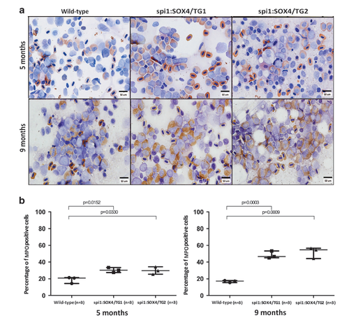
Myeloperoxidase (MPO) staining analysis of spi1:SOX4-EGFP zebrafish. (a) Increased myeloperoxidase was detected in SOX4 transgenic fish at 5 and 9 months (magnification: × 1000). (b) Quantification of MPO-positive cells. Differences among variables were assessed using Student’s t-test. PHENOTYPE:
|
|
Histological analysis of the kidney of Tg(spi1:SOX4-EGFP) zebrafish. H&E stains of the kidney are from 5-, 9- and 12-month-old Tg(spi1:SOX4-EGFP) and wild-type fish. The kidney from spi1:SOX4-EGFP fish showed infiltration by myeloid cells at 9 and 12 -months. Arrow: renal tubule. Yellow triangle: infiltration cells. Magnification: 400x. PHENOTYPE:
|

ZFIN is incorporating published figure images and captions as part of an ongoing project. Figures from some publications have not yet been curated, or are not available for display because of copyright restrictions. PHENOTYPE:
|


