Figure 1—figure supplement 1.

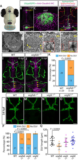
Molecularly heterogeneous networks of the brain and meningeal vasculature and developmental time courses of mCP vascularization.

(A) Dorsal view of a 10 dpf Tg(plvap:EGFP);Tg(glut1b:mCherry) head immunostained for Claudin-5 (magenta) shows heterogeneous Tg(plvap:EGFP) and Tg(glut1b:mCherry) expression levels in the brain and meningeal vasculature. Strong Tg(plvapb:EGFP) and weak Tg(glut1b:mCherry) expression was observed in the mCP vasculature. (B) Dorsal view of a 10 dpf Tg(kdrl:EGFP) larval head immunostained for Claudin-5 (magenta). (C–C") Magnified images of the top boxed area in (B) show Claudin-5+ mesencephalic veins (MsV, yellow arrows). (D–D") Magnified images of the bottom boxed area in (B) show Claudin-5- mCP vasculature composed of the dorsal longitudinal vein (DLV) and posterior cerebral vein (PCeV). (E–H) Dorsal views of 32 (E), 46 (F), 54 (G), and 72 (H) hpf cranial vasculature visualized by Tg(kdrl:EGFP) expression. The DLV began sprouting before 46 hpf (F) and extended posteriorly toward the myelencephalic choroid plexus (mCP) to connect with the bilateral PCeV around 54 hpf (G) to form the mCP vasculature through 72 hpf (H). (I) Schematic representation of the dorsal view of the cranial vasculature at 72 hpf. Scale bars: 50 µm.

Expression Data

Expression Detail
Antibody Labeling
Phenotype Data

Phenotype Detail
Acknowledgments
This image is the copyrighted work of the attributed author or publisher, and ZFIN has permission only to display this image to its users. Additional permissions should be obtained from the applicable author or publisher of the image. Full text @ Elife