FIGURE SUMMARY
Title

Specificity, redundancy and dosage thresholds among gata4/5/6 genes during zebrafish cardiogenesis

Authors
Sam, J., Mercer, E.J., Torregroza, I., Banks, K.M., Evans, T.
Source
Full text @ Biol. Open

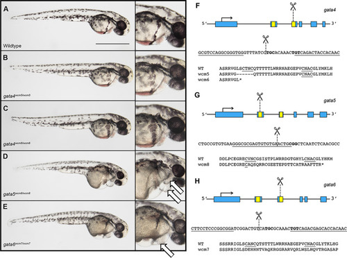
Mutation of gata4 is tolerated while mutationsin gata5 or gata6 cause embryonic lethal cardiac defects. (A–E) Representative images of 2 dpf gata4, gata5, or gata6 homozygous mutants compared to wild type following TALEN (gata4 and gata6) or CRISPR (gata5) mediated deletion of the GATA zinc finger DNA-binding domain. (B,C) Gata4 homozygous mutants are phenotypically normal. (D,E) Gata5 and gata6 homozygous mutants develop severe pericardial edemas (indicated by white arrows in the far right panels) and fail to survive beyond 5–6 days. These phenotypes are 100% penetrant. Representative scale bar in A: 0.5 mm. (F–H) Schematic showing the structure, partial nucleotide sequence, and partial amino acid sequence for each of the gata4/5/6 mutant alleles. Zinc finger domains are shown in yellow and regions targeted for deletion are marked by dashed lines.

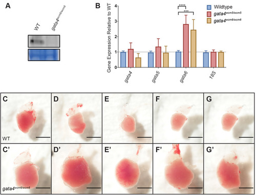
The gata4 mutants are viable to adulthood, but develop an age-dependent cardiomyopathy. (A) Representative western blot showing the absence of Gata4 protein in dissected adult gata4−/− heart tissue. (B) Quantification by qPCR of gata4/5/6 expression reveals a significant upregulation of gata6 mRNA in gata4−/− embryos at the 50% epiboly stage. (C–G) Ventricles dissected from adult wild-type fish, representing phenotypically normal hearts. (C′–G′) Similarly dissected hearts from 10-month-old gata4−/− adults show severe cardiomyopathy and an enlarged ventricular chamber. Scale bar: 1 mm.

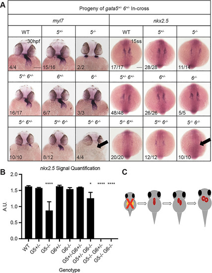
Gata5 and gata6 are redundant for cardiac specification. ISH was performed using offspring from a gata5+/−, gata6+/− in-cross with probes targeting either myocardial marker myl7 (left panels) or cardiac progenitor marker nkx2.5 (right panels). (A) gata5−/−, gata6−/− mutants exhibit a heartless phenotype and a complete loss of cardiac progenitors (indicated by arrows). Representative scale bar in WT panels: 0.1 mm. (B) Quantification of nkx2.5 staining shown in A. Error bars represent s.e.m. *P<0.05, ****P<0.0001. (C) Schematic showing that loss of gata5 and 6 disrupts the initial stages of heart development.

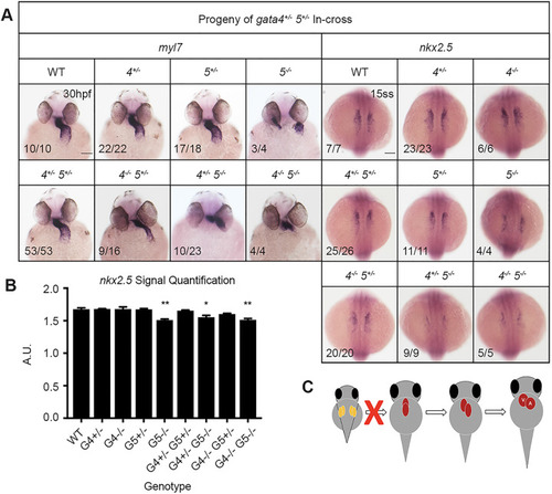
Genetic interactions between gata4 and gata5 impact progenitor migration and heart tube formation. ISH was performed using offspring from a gata4+/−, gata5+/− in-cross and probes targeting either myocardial marker myl7 or cardiac progenitor marker nkx2.5. (A) Progressive loss of gata4 and gata5 alleles results in a range of heart tube defects due to abnormal heart tube morphogenesis. Loss of a gata5 allele in gata4 mutants generates a thin heart tube, while loss of gata4 alleles partially rescues the gata5−/− bifid phenotype. Representative scale bars in WT panels: 0.1 mm. (B) Quantification of nkx2.5 staining shown in A shows that loss of gata4 does not significantly affect cardiac specification. Error bars shown are s.e.m. *P<0.05, **P<0.01. (C) Schematic showing combinatorial loss of gata4 and gata5 disrupts proper fusion of the heart tube.

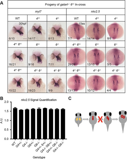
Loss of gata4 and gata6 results in heart tube morphogenesis defects. ISH was performed using offspring from a gata4+/−, gata6+/− in-cross and probes targeting either myocardial marker myl7 or cardiac progenitor marker nkx2.5. (A) The gata4 and gata6 genes are partially redundant for heart tube elongation and looping. The gata4−/−, gata6−/− mutants display distinctly truncated and linear heart tubes. Representative scale bars in WT panels: 0.1 mm. (B) Loss of gata4 and/or gata6 does not affect cardiac specification. Error bars shown are s.e.m. (C) Schematic showing that combinatorial loss of gata4 and gata6 impairs the heart tube elongation stage of cardiogenesis.

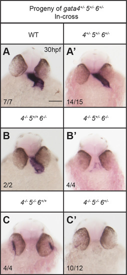
Cardiac development depends on the dosage of gata4/5/6 alleles. ISH was performed using offspring from crossing gata4+/−, gata5+/−, gata6+/− triple heterozygous adults and a probe targeting myocardial marker myl7. (A,A′) Triple heterozygous embryos display abnormal heart tube morphology compared to wild-type siblings, although they can survive to adulthood. Loss of a single gata5 or gata6 allele in a gata4−/−, gata6−/− (B,B′) or gata4−/−, gata5−/− (C,C′) background, respectively, results in a severe loss of cardiac tissue. Representative scale bar in WT panel: 0.1 mm.

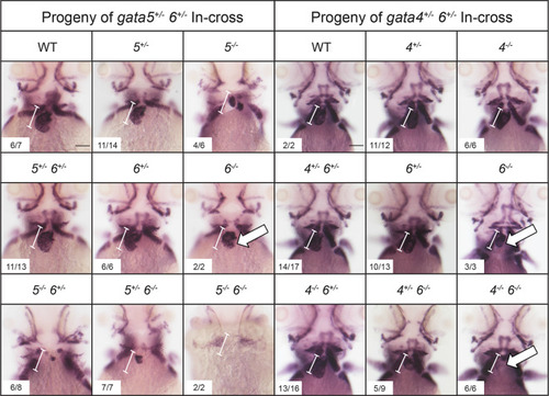
Gata6 regulates ventricular development. ISH was performed at 3 dpf with offspring obtained from crossing either gata5+/−; gata6+/− (left panels) or gata4+/−; gata6+/− (right panels) double heterozygous adult animals with a probe targeting the ventricular marker vmhc. The gata6 homozygous mutants display smaller ventricles (white arrows) compared to the ventricles in wild-type siblings (the normal size of a wild-type ventricle is shown by the bracketed white line, which helps to indicate relative small size of the chamber in mutant embryos). The gata4 null embryos display progressively smaller ventricles with loss of gata6 alleles (white arrows). Representative scale bars in WT panels: 0.1 mm.

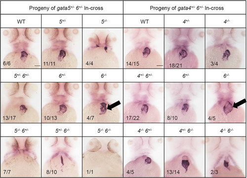
Gata6 mutants exhibit expanded atrial chamber area. ISH was performed at 3 dpf with offspring obtained from crossing either gata5+/−; gata6+/− (left panels) or gata4+/−; gata6+/− (right panels) double heterozygous adult animals with a probe targeting the atrial marker amhc. The gata6 homozygous mutant hearts exhibit increased amhc staining compared to gata4 or gata5 homozygous mutants or wild-type siblings (black arrows). Representative scale bars in WT panels: 0.1 mm.

PHENOTYPE:
Fish:
Observed In:
Stage: Protruding-mouth

Gata6 regulates the number of ventricular cardiomyocytes. Shown are representative images of sibling wild-type (WT) (A) and gata6 null hearts (B) at 2 dpf derived from crossing Tg(myl7:gfp; myl7:dsREDnuc); gata6+/- adults and stained with S46 antibody to distinguish the atrium (blue). Scale bars: 0.1 mm. (C) Quantification of cardiomyocytes in each chamber shows a significant loss of ventricular cardiomyocytes in gata6 homozygous mutants (N=7 per genotype, t-test, P<0.0001, bars shown are s.e.m.). The number of atrial cells in the mutant remains unaffected compared to sibling controls, even though the atrial area is expanded.

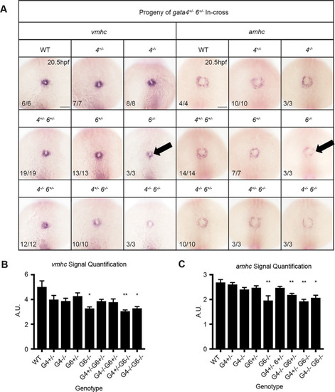
First heart field differentiation is reduced in gata6 mutants. ISH was performed using embryos of the indicated genotypes at 20.5 hpf using probes targeting vmhc or amhc. (A) Ventricular and atrial cardiomyocyte differentiation is decreased in gata6 homozygous mutant embryos (arrows). Representative scale bars in WT panels: 0.1 mm. (B,C) Quantification of vmhc and amhc staining shown in A supports a significant loss of differentiation in gata6 null embryos. Error bars are s.e.m. *P<0.05, **P<0.01.

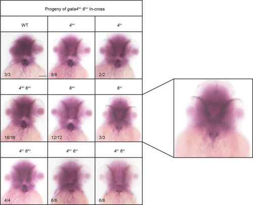
Loss of gata6 impairs second heart field development. ISH was performed using embryos with the indicated genotypes at 2 dpf using a probe targeting second heart field marker ltbp3. The gata6 null embryos express lower levels of ltbp3 compared to gata4 null and wild-type (WT) siblings. Representative scale bar in WT panel: 0.1 mm. Panel on the right is a higher magnification view of representative gata6 mutants.

ZFIN is incorporating published figure images and captions as part of an ongoing project. Figures from some publications have not yet been curated, or are not available for display because of copyright restrictions.

PHENOTYPE:
Fish:
Observed In:
Stage: Long-pec

Unillustrated author statements

PHENOTYPE:
Fish:
Observed In:
Stage Range: Day 5 to Day 6
Acknowledgments
This image is the copyrighted work of the attributed author or publisher, and ZFIN has permission only to display this image to its users. Additional permissions should be obtained from the applicable author or publisher of the image. Full text @ Biol. Open