- Title
-
Zebrafish pax8 is required for otic placode induction and plays a redundant role with Pax2 genes in the maintenance of the otic placode
- Authors
- Mackereth, M.D., Kwak, S.J., Fritz, A., and Riley, B.B.
- Source
- Full text @ Development
|
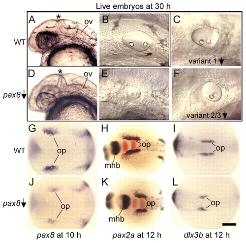
Assessment of Pax8 function in otic induction. (A,D) Head region of live wild-type (A) and pax8-MO injected (D) embryos at 30 hpf. The pax8-MO-injected embryo has a narrow midbrain-hindbrain border (asterisk), and small otic vesicles (ov). (B,E) Otic vesicles at 30 hpf in live wild-type (B) and pax8-MO-injected (E) embryos. (C,F) Otic vesicles at 30 hpf in embryos injected with v1-MO to knockdown variant 1 isoforms of pax8 (C) or v2/3-MO to knockdown variant 2 and 3 isoforms of pax8 (F). (G,J) pax8 expression at 10 hpf in wild-type (G) and pax8-MO-injected (J) embryos. (H,K) Expression of pax2a (blue) and krox20 (red) at 12 hpf in wild-type (H) and pax8-MO-injected (K) embryos. (I,L) dlx3b expression at 12 hpf in wild-type (I) and pax8-MO-injected (L) embryos. Images show lateral views with anterior to the left and dorsal to the top (A-F); or dorsal views with anterior to the left (G-L). mhb, midbrain-hindbrain border; op, otic placode; ov, otic vesicle. Scale bar in L: 200 μm for H,K; 170 μm for G,I,J,L; 150 μm for A,D; 40 μm for B,C,E,F. EXPRESSION / LABELING:
PHENOTYPE:
|
|
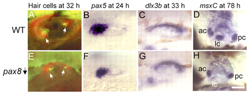
Assessment of Pax8 function in otic vesicle patterning. (A,E) Otic vesicles at 32 hpf stained with anti-Pax2 (red) and anti-acetylated tubulin (green) antibodies to label hair cells (Riley et al., 1999) in wild-type (A) and pax8-MO-injected (E) embryos. Hair cell patches are indicated (white arrows). Injected embryos produce fewer hair cells than normal. In the experiment shown here, an average of 5.2±1.1 hair cells were seen in pax8 morphants (n=59 ears), compared to 9.2±1.4 hair cells in wild type embryos (n=10 ears); 5-10% of pax8 morphants produce only one macula per ear (not shown). (B,F) pax5 expression at 24 hpf in wild-type (B) and pax8-MO-injected (F) embryos. (C,G) dlx3b expression at 33 hpf in wild-type (C) and pax8-MO-injected (G) embryos. (D,H) msxC expression marks developing cristae in the otic vesicle at 78 hpf in wild-type (D) and pax8-MO-injected (H) embryos. The lateral crista is present in the injected embryo but shows strongly reduced expression of msxC. Defects in development of cristae and semicircular canals are highly variable in pax8 morphants. Images show lateral views with anterior to the left and dorsal to the top. ac, anterior crista; lc, lateral crista; pc, posterior crista. Scale bar in H: 90 μm for D,H, 40 μm for C,G, and 30 μm for A,B,E,F. EXPRESSION / LABELING:
PHENOTYPE:
|
|
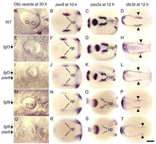
Interaction of pax8 with fgf3 and fgf8. Wild-type embryos (A-D), wild-type embryos injected with fgf3-MO (E-H), wild-type embryos co-injected with fgf3-MO and pax8-MO (I-L), ace (fgf8) mutants (M-P), and ace mutants injected with pax8-MO (Q-T). Images show lateral views of the otic vesicle at 30 hpf (A,E,I,M,Q), and dorsal views of pax8 expression at 10 hpf (B,F,J,N,R), pax2a expression at 12 hpf (C,G,K,O,S) and dlx3b expression at 12 hpf (D,H,L,P,T). op, otic placode. Arrowheads mark the otic region where dlx3b expression normally shows marked upregulation. Anterior is to the left in all specimens. Op, optic placode. Scale bar in T: 30 μm for A,E,I,M; 200 μm for B-D,F-H,J-L,N-P,R-T. EXPRESSION / LABELING:
|
|
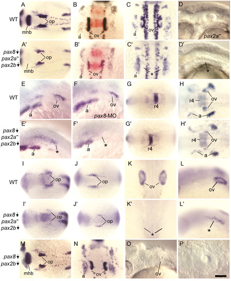
Relative functions of pax8, pax2a and pax2b. (A-C) pax2a expression in wild-type embryos at 14 hpf (A), 18 hpf (B) and 24 hpf (C). (D) Head region of an uninjected noi (pax2a) mutant at 30 hpf. The otic vesicle has essentially normal morphology. A2-D2, noi (pax2a) mutants co-injected with pax8-MO and pax2b-MO showing pax2a expression at 14 hpf (A2), 18 hpf (B2) and 24 hpf (C2), and a live specimen with no otic vesicles at 30 hpf (D2). krox20 expression in the hindbrain is shown in red (B,B2). (E,E2,F,F2) dlx3b expression at 24 hpf in a wild-type embryo (E), a wild-type embryo injected with pax8-MO (F), and noi (pax2a) mutants co-injected with pax8-MO and pax2b-MO (E2,F2). The specimen in E2 lacks morphological otic vesicles but does show a scattering of dlx3b-expressing cells in the otic region (asterisk). The specimen in F2 lacks morphological signs of otic development and also shows no dlx3b expression in the otic region (asterisk). G,G2,H,H2, fgf3 expression in wild-type embryos at 14 hpf (G) and 19 hpf (H) and in pax2ba-pax2b-pax8-deficient embryos at 14 hpf (G2) and 19 hpf (H2). Expression in the hindbrain (r4) is normal in all specimens, whereas expression in the otic vesicle (ov) is strongly reduced at 19 hpf in pax2a-pax2b-pax8-deficient embryos (compare H,H2). (I,I2) sox9a expression at 13.5 hpf in wild-type (I) and pax2a-pax2b-pax8-deficient (I2) embryos. (J-L,J2-L2) claudinA expression in wild-type embryos at 13.5 hpf (J) and 24 hpf (K,L), and in pax2a-pax2b-pax8-deficient embryos at 13.5 hpf (J2) and 24 hpf (K2,L2). M-P, wild-type embryos co-injected with pax8-MO and pax2b-MO showing expression of pax2a at 14 hpf (M) and 18 hpf (N), the head region of a live specimen with a small otic vesicle at 30 hpf (O), and an enlargement of the otic vesicle of the same specimen (P). Images show dorsal views with anterior to the left (A,A2G-J,G2-J2,M), dorsal views with anterior to the top (B,B2,C,C2,K,K2,N), or lateral views with anterior to the left (D-F,D2-F2,L,L2,O,P). a, pharyngeal arches; asterisk, region where otic vesicle normally forms; mhb, midbrain-hindbrain border; op, otic placode; ov, otic vesicle; r4, rhombomere 4 of the hindbrain. Scale bar in P: 170 μm for A,A2,G,G2,I,J,I2,J2,M; 75 μm for B-F,B2-F2,H,H2,K,L,K2,L2,N,O; 19 μm for P. EXPRESSION / LABELING:
|
|
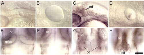
Distinct functions of pax8 splice isoforms. (A-D) Lateral views of the head and otic structures at 30 hpf in noi (pax2a) mutants injected either with pax8 variant 1 MO (A,B) or noi mutants injected with pax8 variant 2/3 MO (C,D). Most noi mutants knocked down for variant 1 isoforms produce a moderate-sized otic vesicle containing hair cells but lacking otoliths (B). In contrast, noi mutants knocked down for variant 2 and 3 isoforms typically produce a small otic vesicle containing both hair cells and otoliths (D) or no otic vesicle at all (data not shown). In addition, all noi mutants knocked down for variant 2 and 3 isoforms show persistent cell death (cd) in the midbrain-hindbrain region. (E-H) Dorsal views of the midbrain-hindbrain border region at 30 hpf in an uninjected wild-type embryo (E), an uninjected noi mutant (F), a noi mutant injected with pax8 variant 1 MO (G) and a noi mutant injected with pax8 variant 2/3 MO (H). Increased cell death is not evident in the majority of noi mutants knocked down for variant 1 isoforms (A) and, if present (G), cell death is diffuse and limited to dorsolateral tissue. In noi mutants knocked down for variant 2 and 3 isoforms, cell death is invariably present, intense, and localized to the midline of the midbrain-hindbrain border region (H). Anterior is to the left (A-D) or to the top (E-F). cd, cell death; mhb, midbrain-hindbrain border; ot, optic tectum. Scale bar in H: 75 μm for A,C; 50 μm for E-H; 19 μm for B,D. PHENOTYPE:
|
|
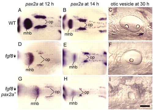
pax2a interacts with fgf8. Wild-type embryos (A-C), ace (fgf8) single mutants (D-F) and ace-noi (fgf8-pax2a) double mutants (G-I). Images show dorsal views of pax2a expression at 12 hpf (A,D,G) and 14 hpf (B,E,H), and lateral views of otic vesicles at 30 hpf (C,F,I). The specimen in B is the same as in Fig. 5A, and the specimen in C is the same as in Fig. 2B. Anterior is to the left in all specimens. mhb, midbrain-hindbrain border; op, otic placode. Scale bar in I: 170 μm for A,B,D,E,G,H; 35 μm for C,F,I. EXPRESSION / LABELING:
PHENOTYPE:
|
|
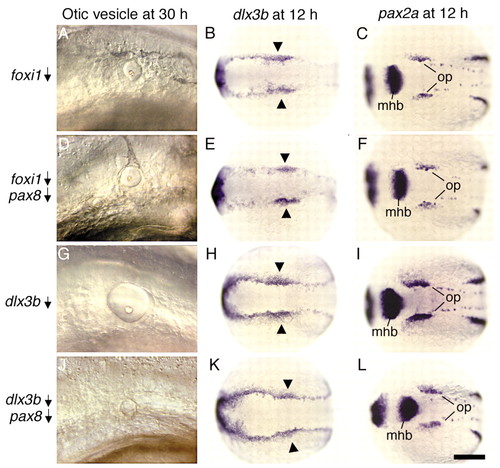
pax8 acts downstream of foxi1 and in parallel with dlx3b. Wild-type embryos injected with foxi1-MO (A-C), foxi1-MO plus pax8-MO (D-F), dlx3b-MO (G-I), or dlx3b-MO plus pax8-MO (J-L). Images show lateral views of the otic vesicle at 30 hpf (A,D,G,J), or dorsal views of dlx3b expression at 12 hpf (B,E,H,K) and pax2a expression at 12 hpf (C,F,I,L). Anterior is to the left in all specimens. mhb, midbrain-hindbrain border; op, otic placode. Arrowheads mark the otic region of dlx3b expression. Scale bar in L: 50 μm for A,D,G,J; 200 μm for B,C,E,F,H,I,K,L. EXPRESSION / LABELING:
PHENOTYPE:
|

Unillustrated author statements |

