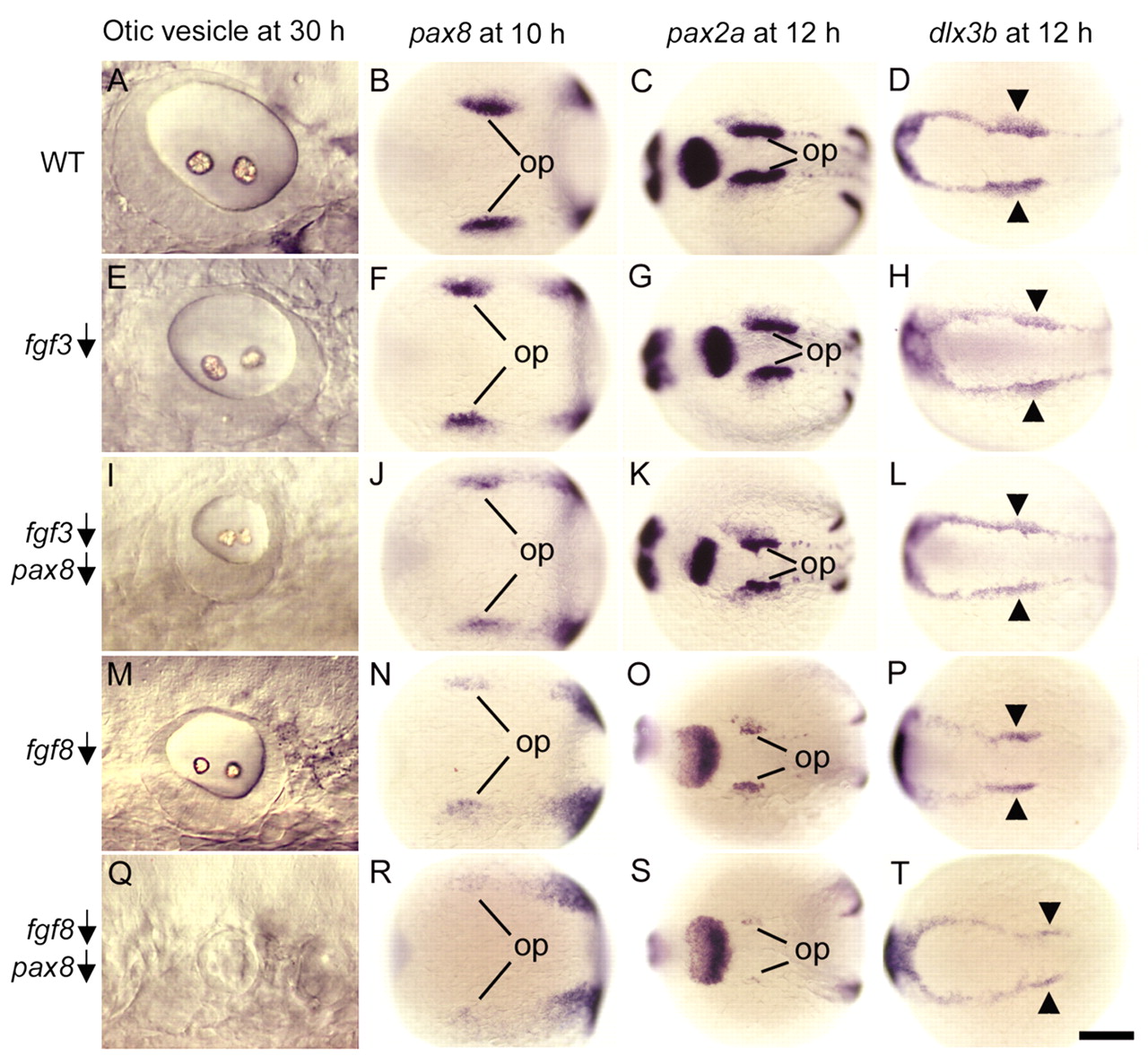
Fig. 4 Interaction of pax8 with fgf3 and fgf8. Wild-type embryos (A-D), wild-type embryos injected with fgf3-MO (E-H), wild-type embryos co-injected with fgf3-MO and pax8-MO (I-L), ace (fgf8) mutants (M-P), and ace mutants injected with pax8-MO (Q-T). Images show lateral views of the otic vesicle at 30 hpf (A,E,I,M,Q), and dorsal views of pax8 expression at 10 hpf (B,F,J,N,R), pax2a expression at 12 hpf (C,G,K,O,S) and dlx3b expression at 12 hpf (D,H,L,P,T). op, otic placode. Arrowheads mark the otic region where dlx3b expression normally shows marked upregulation. Anterior is to the left in all specimens. Op, optic placode. Scale bar in T: 30 μm for A,E,I,M; 200 μm for B-D,F-H,J-L,N-P,R-T.
Image
Figure Caption
Figure Data
Acknowledgments
This image is the copyrighted work of the attributed author or publisher, and
ZFIN has permission only to display this image to its users.
Additional permissions should be obtained from the applicable author or publisher of the image.
Full text @ Development

