
Fig. 2
- ID
- ZDB-FIG-240918-47
- Publication
- Albu et al., 2024 - Distinct mechanisms regulate ventricular and atrial chamber wall formation
- Other Figures
- (all 7)
- All Figure Page
- Back to All Figure Page
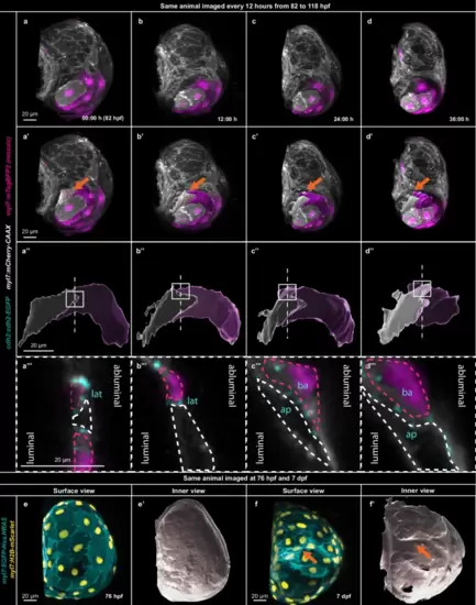
Atrial cardiomyocyte elongation leads to cell intercalation and convergent thickening.a–d 3D airyscan imaging of the same larva every 12 h from 82 to 118 hpf; CM membranes shown in white (myl7:mCherry-CAAX) and mosaic CM cytoplasmic expression in magenta (myl7:mTagBFP2). a’–d” 3D segmentation of two elongating and intercalating CMs reconstructed with opaque (a’–d’) and transparent (a”–d”) surfaces, revealing cell intercalation; orange arrow points to the segmented CMs; squares and dashed lines indicate cross-section region. a”’–d”’, Cross-sections through elongating atrial CMs; dashed lines outline the two intercalating CMs; N-cadherin shown in cyan (cdh2:cdh2-EGFP); lat–lateral adhesion; ap–apical adhesion, ba – basal adhesion; (a–d”’) all 3D surfaces of the mTagBFP2+ CM shown in magenta and all 3D surfaces of the mTagBFP2- CM in white. e, f Outer surface views of 3D airyscan images of the same atrium at 76 hpf and 7 dpf; CM membranes shown in cyan (myl7:EGFP-Hsa.HRAS) and CM nuclei in yellow (myl7:H2B-mScarlet). e’, f’ Inner surface views of the same 3D airyscan images at 76 hpf and 7 dpf; orange arrows point to CMs in the inner ridges. |