|
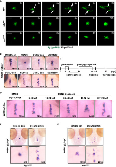
Hgf/met promotes caudal expansion along the pharyngeal midline during the late stage of thyroid development via the MAPK signaling pathway. A The Tg(tg:EGFP) transgenic zebrafish line was used to analyze the time lapse of thyroid development. Representative images chosen from Supplementary Movies 1–3 showing the differential thyroid phenotypes of wild-type and hgfa/met mutant embryos (ventral view, anterior is to the left). Scale bars: 30 μm. White arrows mark that thyroid primordium caudally expand along the pharyngeal midline in wild-type zebrafish. B Thyroid defects in small molecule-treated larvae. Inhibitors of MEK (U0126) successfully mimicked the small thyroid phenotypes in hgfa or met mutant embryos, while inhibitors of MAPK p38 (SB203580), PI3K (LY294002) and STAT3 (SU6656) had little effect on thyroid development. C, D WT embryos were treated with the MEK inhibitor U0126 during the different developmental stage of the zebrafish embryos. In panel C, the treatment periods of gastrulation and somitogenesis cover developmental periods preceding thyroid anlage formation (approximately 24 hpf), whereas the treatment period in the pharyngula represents the developmental period after the onset of thyroid anlage formation. The treatment period from 48–72 hpf covers the developmental period of during which the thyroid primordium forms branches. The treatment period during 3–5 dpf covers the developmental period of thyrocyte proliferation. In panelD, the thyroid primordium was detected by WISH using tg as a probe in 5 dpf larvae after treatment with U0126. Scale bars: 100 μm. E, F The rescue effects of continuously phosphorylated MEK (pTol2tg:pMek) on thyroid development in hgfaK80XE and metI284NF homozygous embryos were visualized via WISH. Scale bars: 100 μm. Three independent experiments were carried for B, D–F.
|