|
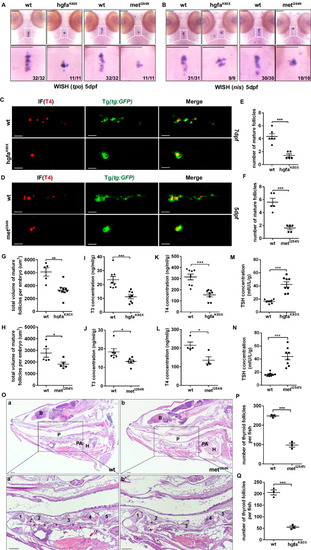
The hgfaK80X and metI284N mutations result in hypothyroidism in zebrafish. A, B The expression of tpo and nis, two specific thyroid markers, in wild-type or homozygous hgfaK80X and metI284N mutant zebrafish larvae at 5 dpf was detected via WISH. C–H Confocal examination of mature thyroid follicles marked by T4 immunofluorescence in the tg transgenic zebrafish line Tg(tg:EGFP) at 7 dpf C and 5 dpf D. C, D: ventral position, anterior is to the left. The total number E, F and volume G, H of thyroid follicles per larva were quantified. n = 6 for WT and n = 5 for hgfaK80X in E (P = 0.0003), n = 5 for WT and n = 5 for metI284N in F (P = 0.00027), n = 5 for WT and n = 9 for hgfaK80X in G (P = 0.0012), n = 5 for WT and n = 5 for metI284N in H (P = 0.0462). I–N The levels of thyroid hormones (T3, T4 and TSH) were detected via ELISA in 1.5-month-old WT and hgfaK80X- or metI284N- mutant zebrafish. The three individual zebrafish were pooled. After homogenization, the T3 and T4 concentrations in the supernatant were measured. Each group was analyzed in triplicates. n = 8 for each group in I (P = 0.00068), K (P = 0.00013), M (P = 0.00002), N (P = 0.00008); n = 6 for each group in J (P = 0.0268); n = 4 for each group in L (P = 0.01999). O Hematoxylin and eosin staining of sagittal sections of thyroid follicles from 1.5-month-old WT and metI284N- mutant zebrafish. P, Q Quantification of the number of thyroid follicles in 1.5-month-old WT and mutant zebrafish. N = 3 for each group (P = 0.0002 for P, P = 0.00026 for Q). B, brain; p, pharyngeal; PA, pericardial aorta; H, heart; 1–5, cartilage; *thyroid follicles. Scale bars: 100 μm. *represents P < 0.05, ***represents P < 0.001. Data are presented as the mean ± SEM. Group comparisons were performed with two-sided Student’s t test. Source data are provided as a Source data file.
|