Figure 8
- ID
- ZDB-FIG-230124-91
- Publication
- Shih et al., 2023 - Identification of the Time Period during Which BMP Signaling Regulates Proliferation of Neural Progenitor Cells in Zebrafish
- Other Figures
- All Figure Page
- Back to All Figure Page
|
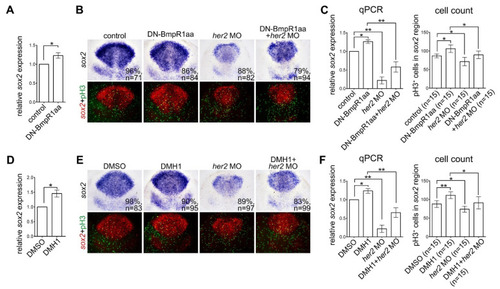
Inactivating BMP signaling upregulates neural progenitor proliferation in response to Her2 knockdown. Heat-shock treatment of Tg (hsp70l:dnBmpr1aa-GFP) or DMH1 was performed at 8–10 hpf embryos and harvested at 10 hpf. (A,D) qRT-PCR analysis demonstrated increased her2 expression by activating DN-Bmpr1aa (A) and DMH1 treatment (D). (B,E) In situ hybridization of sox2 expression demonstrating BMP inactivation by activating DN-Bmpr1aa or DMH1 treatment upregulated sox2 expression and the number of proliferating neural progenitors revealed by sox2 expression and pH3 counterstaining. Injection of her2 morpholino at the one-cell stage significantly reduced sox2 expression and the number of proliferating neural progenitors in embryos with or without BMP inactivation. (C,F) The results of in situ hybridization were confirmed by qRT-PCR, and the number of pH3-positive cells in sox2-expressing areas was counted manually. n, total number of embryos analyzed from three independent experiments. The percentages in each panel in B and E indicate the proportion of embryos displaying the same phenotype as that shown in the photographs of the total embryos examined. Quantitative data are presented as mean ± standard deviation (SD). * p < 0.05; ** p < 0.01. |

