Figure 4
- ID
- ZDB-FIG-230124-87
- Publication
- Shih et al., 2023 - Identification of the Time Period during Which BMP Signaling Regulates Proliferation of Neural Progenitor Cells in Zebrafish
- Other Figures
- All Figure Page
- Back to All Figure Page
|
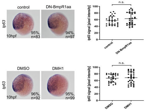
Epidermal ectoderm was unaffected after blocking BMP signaling from 8 hpf. Left panels are lateral views of embryos with ventral to the left. In situ hybridization showed no apparent change in the tp63-expressing region in BMP signal-depleted embryos from 8 hpf onward. n, total number of embryos analyzed from three independent experiments. The tp63 signals were measured using ImageJ and quantified (right panels). n, total number of embryos analyzed from three independent experiments. The percentages in each panel indicate the proportion of embryos displaying the same phenotype as that shown in the photographs of the total embryos examined. Quantitative data are presented as mean ± standard deviation (SD). n.s., not significant. |

