Figure 2
- ID
- ZDB-FIG-230124-85
- Publication
- Shih et al., 2023 - Identification of the Time Period during Which BMP Signaling Regulates Proliferation of Neural Progenitor Cells in Zebrafish
- Other Figures
- All Figure Page
- Back to All Figure Page
|
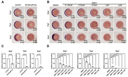
DN-Bmpr1aa and DMH1 sufficiently inhibited expression of eve1 at different time points. All panels are animal pole views with ventral to the left. Expression of eve1 was examined using in situ hybridization. (A) Tg (hsp70l:dnBmpr1aa-GFP) embryos were heat shock-treated at 5.3 hpf and harvested at 6, 7, and 9 hpf showing that eve1 expression was gradually downregulated. (B) Different concentrations of DMH1 were applied to embryos from 5.3 hpf, which inhibited eve1 expression. n, total number of embryos analyzed from three independent experiments. (C,D) qRT-PCR confirmed the expression levels of eve1 of in situ hybridization in (A,B). The percentages in each panel in (A,B) indicate the proportion of embryos displaying the same phenotype as that shown in the photographs of the total embryos examined. Quantitative data are presented as mean ± standard deviation (SD); ****, p < 0.0001. |

