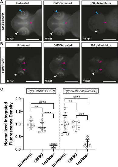
Inhibition of TGFβ signaling with SB431542 reduces pou4f1 enhancer-driven GFP expression in the retina. Representative images of untreated, DMSO-treated and SB431542-treated Tg(12xSBE:EGFP) embryos at 48 hpf (A) showing GFP fluorescence reporting of pSmad3-mediated signaling in the retina (white arrows), brain (magenta arrows) and lens (blue arrows); scale = 250 µm. Representative images of untreated, DMSO-treated and SB431542-treated Tg(pou4f1-hsp70l:GFP) embryos at 48 hpf (B) showing pou4f1 enhancer-driven GFP fluorescence in the retina (white arrows) and optic nerves (magenta arrows); scale = 250 µm. Normalized integrated fluorescence density (C) of each condition (Tg(12xSBE:EGFP); untreated: n = 7, DMSO-treated: n = 7, SB431542-treated: n = 12: ****p < 0.0001, ***p = 0.0004, ns = non-significant; Tg(pou4f1-hsp70l:GFP); untreated: n = 11, DMSO-treated: n = 5 and SB431542-treated: n = 8, ****p < 0.0001, ***p = 0.0004, ns = non-significant; one-way ANOVA Tukey’s multiple comparisons test.
|
