FIGURE

Figure 12

ID
ZDB-FIG-211216-158
Publication
Dalum et al., 2021 - High-Resolution, 3D Imaging of the Zebrafish Gill-Associated Lymphoid Tissue (GIALT) Reveals a Novel Lymphoid Structure, the Amphibranchial Lymphoid Tissue
Other Figures
All Figure Page
Back to All Figure Page
Figure 12

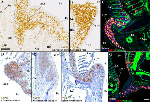
The ALT is a preserved structure between distant teleost species. Representative acquisitions of anti-ZAP70 (brown/red hot) labeled gills section, from different fish species, displaying the side of a gill arch, at the base of filaments. (A, B) ALT of the European carp (Cyprinus carpio); Cyprinid – Paraffin section. (C) ALT of the Atlantic salmon (Salmo salar L.); Salmonid – cryosections stained with phalloidin (green) and DAPI (blue). (D) ALT of the Atlantic mackerel (Scomber scombrus); Scombridae, Percomorph – Paraffin section. (E) ALT of the Northern red snapper (Lutjanus campechanus); Lutjanidae, Percomorph – Paraffin section. (F) ALT of the Pacific barracuda (Sphyraena argentea); Sphyraenidae, Percomorph – Paraffin section. (G) ALT of the Medaka (Oryzias latipes); Adrianichthyidae, Percomorph - cryosections stained with phalloidin (green) and DAPI (blue). These images are maximum intensity projections: 2 µm (G) and 5 µm (C). Annotations: ALT, Amphibranchial Lymphoid Tissue; Bc, Branchial cavity; Bm, Basement membrane; Ea, Efferent artery; Ep, Epithelial cells; F, Filament; Ga, Gill arch; Gr, Gill raker; La, Lamella; S, Septum and Sm, Smooth muscles. Scale bars: 50 µm (B, G) and 70 μm (A, C).

Expression Data

Expression Detail
Antibody Labeling
Phenotype Data

Phenotype Detail
Acknowledgments
This image is the copyrighted work of the attributed author or publisher, and ZFIN has permission only to display this image to its users. Additional permissions should be obtained from the applicable author or publisher of the image. Full text @ Front Immunol