FIGURE

Figure 1

ID
ZDB-FIG-211216-146
Publication
Dalum et al., 2021 - High-Resolution, 3D Imaging of the Zebrafish Gill-Associated Lymphoid Tissue (GIALT) Reveals a Novel Lymphoid Structure, the Amphibranchial Lymphoid Tissue
Other Figures
All Figure Page
Back to All Figure Page
Figure 1

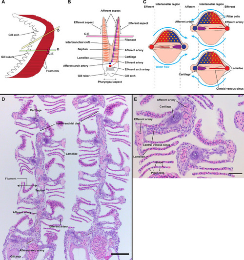
General organization of zebrafish gills. Schematic representations of the overall anatomy of adult zebrafish gills as observed from the side (A), through a transversal plane across the gill arch (B) and through a coronal plane across gill filaments (C). Section plane are illustrated in (A, B). The blood flows from the afferent artery to the lamellae where it gets oxygenated and leave by the efferent artery. Counter-current to the blood, the water irrigates the lamellae from the efferent aspect to the afferent aspect of filaments. (D) Paraffin section (2 µm) displaying two gill arches through an oblique coronal plane, as illustrated in (A). (E) Paraffin section (2 µm) displaying two filaments through a coronal plane, as illustrated in (A, B). Scale bars: 50 µm (D) and 20 µm (E).

Expression Data

Expression Detail
Antibody Labeling
Phenotype Data

Phenotype Detail
Acknowledgments
This image is the copyrighted work of the attributed author or publisher, and ZFIN has permission only to display this image to its users. Additional permissions should be obtained from the applicable author or publisher of the image. Full text @ Front Immunol