FIGURE

Figure 6

ID
ZDB-FIG-211216-152
Publication
Dalum et al., 2021 - High-Resolution, 3D Imaging of the Zebrafish Gill-Associated Lymphoid Tissue (GIALT) Reveals a Novel Lymphoid Structure, the Amphibranchial Lymphoid Tissue
Other Figures
All Figure Page
Back to All Figure Page
Figure 6

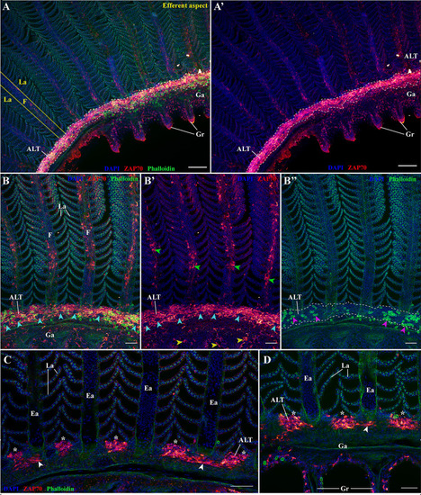
The amphibranchial lymphoid tissue is a continuous structure along gill arches. Representative deconvolved confocal images of an adult zebrafish gill arch observed from the side. The images were acquired from wholemount dissected gill arches stained with phalloidin (green) and DAPI (blue) and where T/NK cells were labeled with anti-ZAP70 antibody (red hot). (A,A’) low magnification image of the gill arch illustrating the continuity of the ALT. (B–B’’) Images of the ALT at a higher magnification showing the contrast between the lymphoid organization of the ALT, the efferent aspect of filaments (green arrowheads), and the pharyngeal side of the gill arch (yellow arrowheads). Magenta arrowheads in (B’’) emphasize the enrichment of the ALT in rodlet cells, while the cyan arrowheads in (B, B’) point to the localization of neuromast-like ZAP70 negative structures. (C, D) Optical sections from within 3D acquisitions illustrating the variating thickness of the ALT. The white arrowheads point to the thinness of the ALT when at the level of the efferent artery compared to the thickness of the ALT when in-between filaments. (D) is deeper within the 3D acquisition than (C). (A–D) Images are maximum intensity projections: 5 μm (C, D), 25 μm (B–B’’) and 50 μm (A, A’). Annotations: ALT, Amphibranchial Lymphoid Tissue; Ea, Efferent artery; F, Filament; Ga, Gill arch; Gr, Gill raker and La, Lamellae. Scale bar: 20 μm (D), 40 μm (B, C) and 100 μm (A, A’).

Expression Data

Expression Detail
Antibody Labeling
Phenotype Data

Phenotype Detail
Acknowledgments
This image is the copyrighted work of the attributed author or publisher, and ZFIN has permission only to display this image to its users. Additional permissions should be obtained from the applicable author or publisher of the image. Full text @ Front Immunol