|
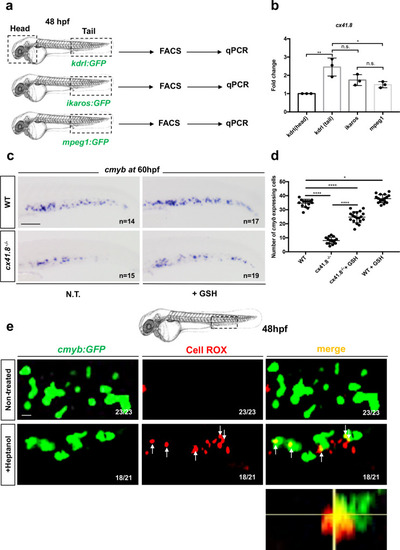
<italic>Cx41.8</italic><sup><italic>−/−</italic></sup> mutant embryos show a decrease in HSPCs which is rescued by antioxidant treatment.a qPCR data examining cx41.8 expression (fold change relative to expression in kdrl:GFP+ head subset) in FACS-sorted cells from kdrl:GFP, ikaros:GFP and mpeg1:GFP embryos. GFP+ cells from dissected heads and tails were sorted from 48 hpf embryos. b For each transgenic line have been pooled n = 20 animals, and each experiment was repeated independently three times. Statistical analysis was completed using a one-way ANOVA, with Tukey–Kramer post hoc tests, adjusted for multiple comparisons *P = 0.018; **P = 0.001; (n.s.) non-significant P = 0.26; P = 0.74. ccmyb expression at 60 hpf in wild type and cx41.8−/− embryos, and after GSH treatment. d Quantification of cmyb-expressing cells. Statistical analysis: one-way ANOVA, with Tukey–Kramer post hoc tests, adjusted for multiple comparisons, *P = 0.045; ****P < .0001. Center values denote the mean, and error values denote s.e.m. e Imaged area in the tail at 48 hpf, as indicated by the black dotted line. Confocal imaging of the CHT at 48 hpf of cmyb:GFP+ cells (green), CellROX fluorescent probe (red) and merge, in NT and after heptanol treatment cmyb:GFP embryos. Orthogonal projection CellROX+cmyb:GFP+ double-positive cell after heptanol treatment. Scale bars: 100 μm (c); 25 μm (e).
|

