|
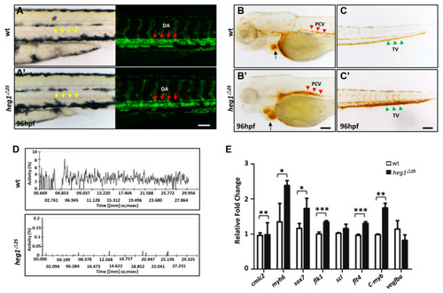
heg1 deficiency leaded to poor blood flow and abnormal vascular development in zebrafish embryos. (A) Lateral view of zebrafish larvae at 96 hpf. Representative images of the heg1∆25 and wt embryos, exhibiting blood congestion (yellow arrows), and dilation of dorsal aorta (DA) lumen (red arrows) in caudal vein. (B,C) Representative images of the heg1∆25 and wt embryos, showing red blood cells (RBCs) accumulation in posterior cardinal vein (PCV) (red arrowhead), tail vein (TV) (green arrowhead) and heart (black arrow). (D) The movement ratio of RBCs based on changes in pixel density of PVC. (E) The expressions of cardiovascular markers, as determined by qRT-PCR, were significantly changed in heg1∆25 mutants at 48 hpf. Data are represented as mean ± SE from three independent experiments, * p < 0.05, ** p < 0.01, and *** p < 0.001 (Student’s t-test).
|