|
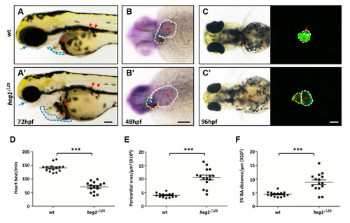
heg1 deficiency leaded to abnormal cardiac development in zebrafish embryos. (A,A’) Lateral view of zebrafish larvae at 72 hpf. Representative images of the heg1∆25 and wt embryos, exhibiting pericardial edema (blue dotted-line boxes), enlarged heart size (green arrow), venous congestion (red arrow), and eye edema (blue arrow). (B,B’) Representative images of the heg1∆25 and wt embryos at 48 hpf stained for the heart marker cmlc1. Note the enlargement heart in heg1∆25 mutants (V: Ventricular, yellow dotted-line boxes; A: atria, white dotted-line boxes, ventral view). (C,C’) The heart morphology was delineated by Tg(cmlc2:GFP) (ventral view). (D) Heart rate in wt and heg1∆25 mutant zebrafish larvae (n = 15 embryos/group). (E) The pericardial area in wt and heg1∆25 mutant zebrafish larvae (n = 15 embryos/group). (F) The SV-BA distance in wt and heg1∆25 mutant zebrafish larvae (n = 15 embryos /group). Scale bar: 100 μm. *** indicates p < 0.001 by Student’s t-test.
|