
Fig. 3
- ID
- ZDB-FIG-160224-3
- Publication
- Ulrich et al., 2016 - Reck enables cerebrovascular development by promoting canonical Wnt signaling
- Other Figures
- All Figure Page
- Back to All Figure Page
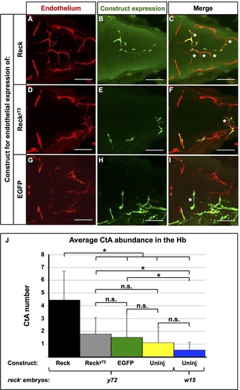
Mosaic endothelial expression of WT Reck is sufficient to rescue the CtA deficit of reck y72 mutant embryos. (A-I) Dorsal views (dorsal level to show CtAs) of the 72hpf Hb vasculature {red [Tg(kdrl:RFP)s896]} of reck y72 injected with constructs driving endothelial expression of exogenous Reck, Recky72 (both HA-tagged, see Fig. 2L) or EGFP proteins (green). Anterior, left; right side, up. (C,F,I) White asterisks indicate CtAs with exogenous expression of listed proteins. Scale bars, 100µm. (J) Quantification of CtA abundance in the Hb of reck y72 and Df(Chr24:reck)w15 with or without (‘Uninj’) exogenous endothelial expression of listed proteins. Asterisks indicate significant differences (P<0.001); n.s., not significant; Student′s t-test. reck y72 mutants scored: Reck (n=28), Recky72 (n=14), EGFP (n=19), Uninj (n=20). Df(Chr24:reck)w15 mutants scored: Uninj (n=19). See also Fig. 4, Figs S6, S11. |