
Fig. S7
- ID
- ZDB-FIG-160224-13
- Publication
- Ulrich et al., 2016 - Reck enables cerebrovascular development by promoting canonical Wnt signaling
- Other Figures
- All Figure Page
- Back to All Figure Page
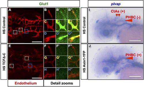
Forced inhibition of canonical Wnt signaling impairs CtA formation and leads to aberrant cerebrovascular expression of the Wnt-responsive markers of barriergenic differentiation Glut1 and plvap. (A-H′′) Confocal dorsal images of the 48 hpf Hb vasculatures of heat-shocked (HS) embryos without (Control) or with TCFΔC (Tg(hsp70l:Xla.TCFΔC-EGFP) over-expression. Heat-shock: 40°C for 30 minutes at 30 hpf. Anterior, left. Right side, up. Endothelium, red (Tg(kdrl:RFP)s896); Glut1 immunofluorescence, green. Colored dashed boxes (A, E) demarcate a region of the following vessels: white, MtA (zooms: B-B′′, F-F′′); yellow, CtAs (zooms: C-C′′, G-G′′); blue, PHBCs (zooms: DD′′, H-H′′). Merged images of zooms: B′′, C′′, D′′, F′′, G′′, H′′. Glut1 decorates the MtA, CtAs and PHBCs of the Control (n=2 embryos). In contrast, in the TCFΔC over-expressing embryo CtA abundance is reduced (n=10 embryos) and cerebrovascular Glut1 decoration is missing (n=2 embryos). (I-J) Transmitted-light lateral images of the 48 hpf heads of heat-shocked (HS) embryos without (Control) or with Axin1-YFP (Tg(hsp70l:Mmu.Axin1-YFP)w35) over-expression subjected to whole mount RNA in situ hybridization with plvap riboprobes. Heat-shock: 39oC for 1 hour at 24 hpf. (I) Control. plvap is expressed in the dorsal aspect of CtAs but not in the PHBCs (n=9 embryos). (J) Axin1-YFP overexpressing embryo. plvap is ectopically expressed in the PHBCs (n=7 embryos). Anterior left. Dorsal up. Scale bar: 100 µm. |