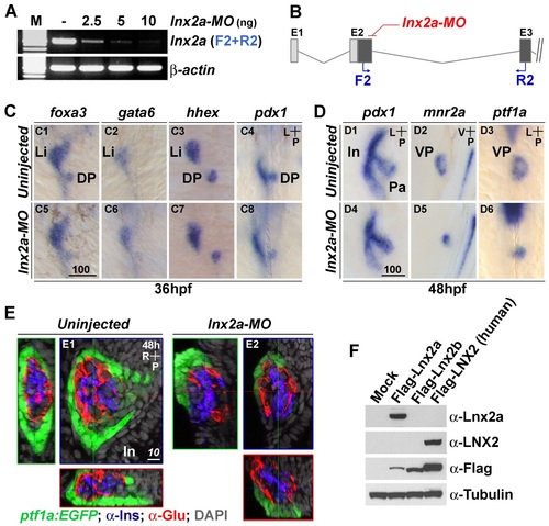
lnx2a knock-down affects development of the exocrine pancreas. (A) RT-PCR reveals that lnx2a-MO injection efficiently blocks pre-mRNA splicing between exon 2 and exon 3. (B) A schematic drawing indicates the location of primers in exon 2 (F2) and exon 3 (R2). (C and D) lnx2a is involved in the maintenance of ventral pancreas but not initial establishment of endoderm patterning. (C) At 36 hpf, foxa3, gata6, hhex and pdx1-positive cells develop normally in lnx2a-MO injected embryos. (D) At 48 hpf, pdx1-expressing pancreas precursor tissue is normally formed, but mnr2a and ptf1a-postive ventral pancreas precursors are reduced in lnx2a- MO injected embryos (D5 and D6). (E) Detection of lnx2a-MO effect using transgenic embryos and immuno staining. Tg(ptf1a:EGFP) embryos were injected with lnx2a-MO and stained for insulin (Ins) and glucagon (Glu) to visualize β-cells and α-cells, respectively. Confocal images at 48 hpf are shown along three axes. lnx2a-MO injected embryos display defective ventral pancreas (ptf1a: EGFP+), but normal dorsal pancreas-derived β-cells (Ins+) and α-cells (Glu+). (F) Western blot shows the specificity of the anti-Lnx2a polyclonal antibody. Flag-tagged zebrafish Lnx2a, Lnx2b and human LNX2 expression vectors were transfected into 293T cells, and lysates were analyzed by immuno blotting with anti-Lnx2a, LNX2, Flag and Tubulin antibodies, as indicated. Li, Liver; Pa, Pancreas; In, Intestine; DP, Dorsal pancreas; VP, Ventral pancreas; Scale bar, 100 µm (C5) and 10 µm (E1).
|

