|
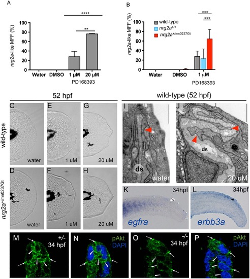
The nrg2a MFF phenotype can be mimicked and synergistically enhanced via chemical ErbB inhibition, and is characterized by reduced pAKT levels. (A-J) Pharmacological inhibition of ErbB signaling during MFF morphogenesis (24 through 52 hpf) induces nrg2amn0237Gt/mn0237Gt-like effects in wild-type and, with even higher frequencies, nrg2a+/mn0237Gt heterozygous embryos. (A) MFFs of wild-type embryos treated both with a low dose (1 µM) (28%; n = 120; p = 0.005) or a high dose (20 µM) (76%; n = 102; p < 0.0001) of PD168393 show an nrg2amn0237Gt/mn0237Gt-like MFF morphology at 52 hpf. (B) nrg2a+/mn0237Gt heterozygous embryos (red) are significantly more sensitive to low-dose (1 µM) PD168393 treatment, and display nrg2amn0237Gt/mn0237Gt-like MFF morphology with a higher frequency (64%; n = 152) than treated nrg2a+/+ wild-type siblings (blue) (n = 23%; n = 116; p < 0.0001) or treated unrelated wild-type embryos (grey) (28%; n = 120; p < 0.0001). (D-H) Live images of MFFs of representative examples of wild-type (C, E, G) or nrg2a+/mn0237Gt heterozygous (D, F, H) embryos at 52 hpf, after treatment with DMSO (control; C, D), 1 µM PD168393 (E, F) or 20 µM PD168393 (G, H). (I, J) TEM transverse sections of the apical MFF reveal a ridge cell phenotype in PD168393-treated wild-type embryos at 52 hpf. (I) An untreated wild-type embryo has correctly elongated ridge cells (red arrowhead) and a straight dermal space (ds). (J) A sibling embryo treated with 20 µM PD168393 displays basal bulging of MFF ridge cells towards the center the fin fold (red arrowheads) and a corresponding serpentine-like folding of the dermal space (ds), resembling the defects of the nrg2a mutant (compare with Fig 8D). (K, L) Whole-mount in situ hybridization (WISH) for egfra (K) and erbb3a (L) in wild-type embryos at 34 hpf (lateral views of tail) reveals epidermally expressed erbb3a transcripts (L), whereas egfra transcipts are absent in the epidermis, but present in the somites (K). (M-P) Anti-pAKT immunofluorescence of wild-type (M, N) and nrg2a mutant (O, P) embryo at 34 hpf; transverse sections through MER region of MFF, counterstained with DAPI (N, P). The wild-type embryo (M, N) displays pAKT localization in distal epidermal MFF cells (ridge cells and cleft cells; arrowheads), whereas pAKT levels in more proximal epidermal MFF cells are much lower. In addition, pAKT is localized at the tight junctions of the outer EVL (arrows), consistent with previously described roles of pAKT to phosphorylate tight junction proteins ZO-1 and Occludin, and to tighten the junctions [122]. In the nrg2a mutant (O, P), pAKT signals in ridge and cleft cells are strongly reduced (arrowheads), while pAKT signals at EVL tight junctions are unaltered (arrows).
|

