|
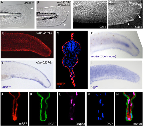
nrg2a mutants display altered MFF morphology, consistent with the epidermal localization of the Nrg2-mRFP fusion protein and of endogenous nrg2a transcripts. (A, B) By 48 hpf, nrg2amn0237Gt/mn0237Gt mutants (mn0237Gt/mn0237Gt) show altered MFF morphology. (A) Wild-type MFF edges are thin, flat, and continuously curved (arrowhead). (B) mn0237Gt/mn0237Gt mutant MFFs have thickened edges (arrowhead), and one or more pointed protrusions (open arrowhead). (C, D) Collagen II (Col II) immunostaining of actinotrichia support fibers within the MFF shows aberrant collagen accumulation and ectopic actinotrichia within mn0237Gt/mn0237Gt mutant apical MFFs (arrowheads) at 48 hpf. (E, G) At 24 hpf, Nrg2a-mRFP fusion protein is present in MFFs of heterozygous (+/mn0237Gt) embryos (E; view on tail of whole mount) and, at slightly lower levels, throughout the entire epidermis (G; section through tail region; immunostained for RFP and counterstained with DAPI). (F, H, I) Whole-mount in situ hybridization (WISH) demonstrates strong MFF expression of the GBT-generated fusion transcript (mRFP; F) in a representative +/mn0237Gt embryo at 24 hpf. When developed with Boehringer Blocking Reagent, WISH staining for endogenous nrg2a transcripts in 24 hpf wild-type embryos also revealed strong MFF expression of the endogenous gene (H, Boeringer). When developed without Boehringer Blocking Reagent, WISH staining further reveals uniform expression of the endogenous nrg2a gene throughout the entire epidermis (I). For cross-sections, see Honjo et al. (2008), Fig 6C [51]). (J-N) Co-labeling of a transverse section through the dorsal MFF of a +/mn0237Gt embryo at 24 hpf reveals restricted localization of the Nrg2a-mRFP fusion protein (J) in &916;Np63-positive basal keratinocytes (L), whereas the outer enveloping layer, labeled with EGFP (K), lacks the Nrg2a-mRFP protein; (M) DAPI counterstain; (N) merged image of different channels shown in (J-M).
|

