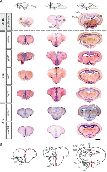
Fig. 5
- ID
- ZDB-FIG-130906-12
- Publication
- Weger et al., 2013 - Real-time in vivo monitoring of circadian E-box enhancer activity: A robust and sensitive zebrafish reporter line for developmental, chemical and neural biology of the circadian clock
- Other Figures
- All Figure Page
- Back to All Figure Page
|
Luciferase expression overlaps with various neurogenic areas and matches clock gene expression throughout the brain. (A) Comparison of luciferase and endogenous clock gene in situ hybridization on adult brain sections. Rows show in situ hybridizations with antisense probes for the indicated genes and timepoints. Columns display brain sections at the level of the telencephalon (left), diencephalon (middle), and mesencephalon (right). Top row: schematic lateral views of the adult zebrafish brain indicating the section level. Luciferase transcripts are detected in the dorsal telencephalic area (D), and in the ventral (Vv), the dorsal (Vd) and the central (Vc) nuclei of the ventral telencephalon. More posteriorly, reporter expression is detected in the preoptic area (PPa), the posterior post-commissural nucleus of the ventral telencephalon (Vp), and in the dorsomedian (Dm) and dorsolateral (Dl) telencephalon. In more caudal sections, luciferase transcripts are detected in the periglomerular gray zone (PGZ) of the optic tectum (TeO), the valvula of the cerebellum (VCe), the torus longitudinalis (TL), the corpus mamillare (CM) as well as in the ventral (Hv) and dorsal (Hd) zones of the periventricular hypothalamus. Expression of all the examined endogenous clock genes shows extensive overlap with luciferase expression. (B) Schematic indication of anatomical subdomains at the levels of the sections in (A) (modified from Wullimann et al. (1996)). Indicated in red are zones of proliferation. Scale bar, 250 μm. |
| Genes: | |
|---|---|
| Fish: | |
| Condition: | |
| Anatomical Terms: | |
| Stage: | Adult |
Reprinted from Developmental Biology, 380(2), Weger, M., Weger, B.D., Diotel, N., Rastegar, S., Hirota, T., Kay, S.A., Strähle, U., and Dickmeis, T., Real-time in vivo monitoring of circadian E-box enhancer activity: A robust and sensitive zebrafish reporter line for developmental, chemical and neural biology of the circadian clock, 259-73, Copyright (2013) with permission from Elsevier. Full text @ Dev. Biol.

