Fig. 5
- ID
- ZDB-FIG-120315-77
- Publication
- Shen et al., 2012 - The cytokine macrophage migration inhibitory factor (MIF) acts as a neurotrophin in the developing inner ear of the zebrafish, Danio rerio
- Other Figures
- All Figure Page
- Back to All Figure Page
|
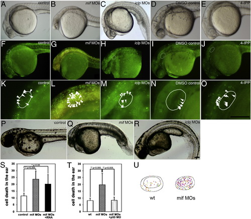
Gross morphology was affected by mif and iclp morpholinos. Lateral view of embryos at 29 hpf (A–E) and 48 hpf (P–R). (F–O) Acridine orange (AO) staining demonstrated cell death in 29-hpf embryos. (K–O) Magnified ear area in (F–J) respectively. (A, F, K, P) control; (B, G, L, Q) mif morphants; (C, H, M, R) iclp morphants; (D, I, N) DMSO control; (E, J, O) 4-IPP (40 μM)-treated embryos. White oval outlines indicate the positions of the otocysts; white arrowheads point to the AO-stained cells in the otocysts. (S, T) Comparison of AO-stained cells in the inner ear (n = 4 for each treatment in (S), and n = 5 for wt, 8 for both mif MOs and mif MOs + p53 MO in (T)) at 29 hpf. (U) Illustration of the location of AO-positive cells in the otocyst. The different colors of the spots represent AO-positive cells in the different embryos assessed. They are superimposed in this figure to determine if cells in a particular region were more susceptible (n = 5 for wild type, and 8 for mif morphants). Scale bar: 100 μm. |
| Fish: | |
|---|---|
| Knockdown Reagents: | |
| Observed In: | |
| Stage Range: | Prim-5 to Long-pec |
Reprinted from Developmental Biology, 363(1), Shen, Y.C., Thompson, D.L., Kuah, M.K., Wong, K.L., Wu, K.L., Linn, S.A., Jewett, E.M., Shu-Chien, A.C., and Barald, K.F., The cytokine macrophage migration inhibitory factor (MIF) acts as a neurotrophin in the developing inner ear of the zebrafish, Danio rerio, 84-94, Copyright (2012) with permission from Elsevier. Full text @ Dev. Biol.

