|
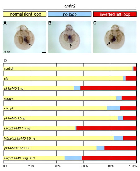
PCP signalling regulates heart loop laterality in zebrafish. (A-C) Top view of 36 hpf pk1aMO-injected embryos showing expression of cardiac myosin light chain 2 (cmlc2) in the heart tube by wholemount in situ hybridisation. Arrows indicate the orientation of heart looping: normal loop to the right (A), absent loop (B) and inverted loop to the left (C). (D) Horizontal bar chart of the relative frequency of heart loop orientation (right loop, yellow; no loop, blue; left loop, red) scored in different experimental conditions (same conditions as in Fig. 1): control (n=122); slb (n=12); pk1a-MO 3 ng (n=137); MZppt (n=122); slb;ppt (n=50); pk1a-MO 1.5 ng (n=94); slb;pk1a-MO 1.5 ng (n=170); MZppt;pk1aMO 1.5 ng (n=60); pk1a-MO 3 ng DFC (n=46); and slb;pk1a-MO 4 ng DFC (n=56). Scale bar: 20 μm.
|