
Fig. S1
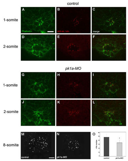
Cilia formation is impaired in pk1a-MO embryos. Confocal images of the dorsal forerunner cell (DFC) cluster double-labelled with the F-actin marker Phalloidin (green) and an anti-acetylated α-tubulin antibody (red). Single dorsal focal planes (individual and merged channels) are shown. (A-F) Control embryos. One-somite stage; DFCs are positioned around a single small lumen that is delineated by strong Phalloidin labelling (A,C) and short acetylated α-Tubulin-rich cilia (B,C). Two-somite stage; a larger lumen and slightly longer cilia are present (D-F). (G-L) Embryos injected with pk1a-MO. One-somite stage; two Phalloidin-rich focal points but no clear lumen are present in the DFC cluster (G,I). Dot-like acetylated α-Tubulin label indicates the initiation of cilia at the position of the focal points (H,I). Two-somite stage; a small mis-shapen lumen strongly labelled by Phalloidin is present (J,L). The lumen is lined with abnormally short cilia (K,L). (M,N) 3D projections of anti-α-Tubulin staining of the Kuppfer′s vesicle (KV) in an 8-somite stage control (M) and pk1a-MO-injected (N) embryos. (O) Cilia number is reduced in pk1a-MO embryos compared with controls (mean ± SD; n=7 control embryos and 7 morphants). Scale bars: 20 μm. |