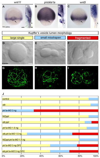
The PCP signalling components wnt11 and prickle1a are expressed in the DFCs and control KV morphogenesis. (A-C′) Expression of wnt11, pk1a and wnt5 in wild-type zebrafish embryos at 75% epiboly as revealed by wholemount in situ hybridisation. Arrows in dorsal views (A,B,C) and arrowheads in sagittal sections (A2,B2,C2) point to the dorsal forerunner cell (DFC) expression domain. (D-I) Kuppfer′s vesicle (KV) lumen morphologies at the 8- to 10-somite stage in wild-type (D,G) and wnt11- and pk1a-defective (E,F,H,I) embryos. (D-F) DIC microscopy views of KV in living embryos. (G-I) Three-dimensional projections of confocal stacks through the entire KV stained with anti-ZO-1 antibody, outlining lumen shape. (J) Horizontal bar chart of the relative frequency of the three main KV morphological phenotypes (large single lumen, yellow; small mis-shapen lumen, blue; fragmented lumen, red) scored in different experimental conditions: control (wild-type untreated, n=21); slb [wnt11 (silberblick) mutants, n=12]; pk1a-MO 3 ng (wild-type injected with 3 ng of pk1a-MO at the 1-cell stage, n=9); MZppt [maternal zygotic wnt5 (pipetail) mutants, n=10]; slb;ppt (wnt11;slb and wnt5;ppt double mutants, n=14); pk1a-MO 1.5 ng (wild-type injected with 1.5 ng of pk1a-MO at the 1-cell stage, n=8); slb;pk1a-MO 1.5 ng (slb mutants injected with 1.5 ng of pk1a-MO at the 1-cell stage, n=19); MZppt;pk1a-MO 1.5 ng (MZppt injected with 1.5 ng of pk1a-MO at the 1-cell stage, n=10); pk1a-MO 3 ng DFC [abrogation of pk1a in wild-type DFCs by injection of 3 ng of pk1a-MO in the yolk syncitial layer (YSL) at mid-blastula stages, n=18]; and slb;pk1a-MO 3 ng DFC (abrogation of pk1a in slb DFCs by injection of 3 ng of pk1a-MO in the YSL at midblastula stages, n=10). Scale bars: 20 μm.
|
