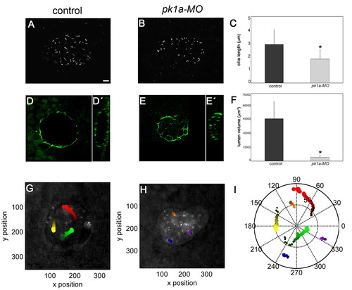
PCP signalling regulates cilia length, lumen volume and Nodal flow in the KV. (A-C) Comparison of cilia length of control and pk1a-MO-injected 8-somite stage zebrafish embryos. z-projections of a dorsal confocal stack through the KV of a control (A) and a morphant (B) embryo stained with an anti-acetylated α-tubulin antibody reveal a more unevenly shaped lumen and shorter cilia in the morphant. Quantitative analysis of multiple embryos (C) reveals significantly shorter cilia in pk1a-MO embryos compared with controls (mean ± s.d.; n=5 control embryos and 3 morphants; *, P<0.05, Student′s t-test). (D-E′) Anti-ZO-1 staining of the KV in control and pk1a-MO-injected embryos. Single dorsal focal planes at the centre of the vesicle (D,E) and sagittal views (D′,E′) are shown. (F) KV lumen volume is reduced in pk1a-MO embryos compared with controls [mean ± s.d.; n=10 control embryos and 5 morphants; *, P<0.05, Student′s t-test (KaleidaGraph, SynergySoftware)]. Approximate volume was determined based on the three main axes of the ellipsoid lumen. For fragmented lumina, volume was computed as the sum. (G-I) Nodal flow is strongly reduced in pk1a-MO embryos. Colour-coded tracks of fluorescent beads injected into the KV of a control (G) and a pk1a-MO (H) embryo. The bead trajectories shown in G and H are plotted in I. Scale bars: 20 μm.
|
