FIGURE SUMMARY
Title

FARS2 Deficiency Causes Cardiomyopathy by Disrupting Mitochondrial Homeostasis and the Mitochondrial Quality Control System

Authors
Li, B., Liu, F., Chen, X., Chen, T., Zhang, J., Liu, Y., Yao, Y., Hu, W., Zhang, M., Wang, B., Liu, L., Chen, K., Wu, Y.
Source
Full text @ Circulation

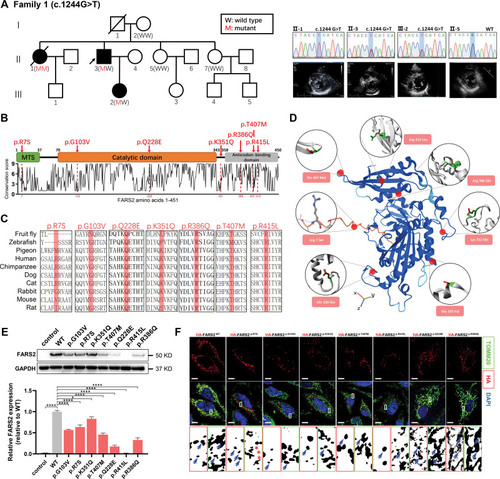
Functional characterization of FARS2 variants identified in hypertrophic cardiomyopathy. A, Pedigrees. Sanger validation of FARS2 variant and echocardiography of the patients in family 1. B, A graphical illustration of FARS2 sequence conservation (bottom) based on ConSurf conservation score. The domain structure and positions of identified patient variants (red symbols) are indicated. C, Species conservation of FARS2 (mitochondrial phenylalanyl-tRNA synthetase) amino acids p.R7, p.G103, p.Q228, p.K351, p.R386, p.T407, and p.R415. D, A structural model of wild-type (WT) human FARS2 (AlphaFold Protein Structure Database O95363 [SYFM_HUMAN]), highlighting the sites of FARS2 variants (p.Arg7Ser, p.Gly103Val, p.Gln228Glu, p.Lys351Gln, p.Arg386Gln, p.Thr407Met, and p.Arg415Leu). E, The expression level of FARS2 from HeLa cells transfected with empty vector control, WT, or 7 variant vectors. The relative statistical analysis is shown in the bottom panel (n=3 per group). F, Confocal images of HeLa cells transfected with HA-FARS2 constructs and immunolabeled with TOMM20 (mitochondrial marker). The regions were magnified with split channels beneath each group and boxed with red (HA) or green (TOMM20). The representative colocalization parts are emphasized by blue arrows; the noncolocalization parts are highlighted by red asterisks. Scale bar=10 μm. *P<0.05; ****P<0.0001.

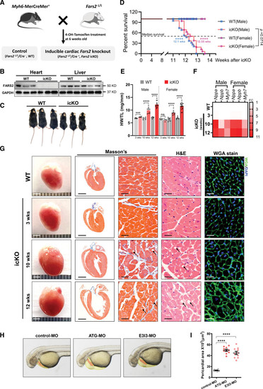
FARS2 deficiency causes cardiac hypertrophy leading to heart failure and sudden death in vivo. A, Schematic diagram to generate 4-OH tamoxifen inducible cardiac-specific Fars2 knockout (icKO) mice by crossing Fars2LoxP/LoxP mice with Myh6-MerCreMer+ mice. B, Western blot analysis of FARS2 (mitochondrial phenylalanyl-tRNA synthetase) protein levels in the heart and liver of wild-type (WT) control and FARS2 icKO mice, with GAPDH as loading control. C, Photograph of WT and icKO male mice at 12 weeks after icKO. D, Kaplan-Meier survival curves for WT and icKO mice (n=16–19). The median survival times for the icKO group are marked. E, Ratios of heart weight to tibia length (HW/TL; n=3–16). F, Relative mRNA levels of cardiac hypertrophy markers (Nppa, Nppb, and Myh7) in mice 3, 10, and 12 weeks after icKO (n=3). G, Representative heart (scale bar=2 mm), longitudinal sections (scale bar=2 mm), Masson trichrome staining (scale bar=25 μm), hematoxylin & eosin staining (scale bar=25 μm), and wheat germ agglutinin staining (scale bar=25 μm) from male mice at 3, 10, and 12 weeks after icKO and WT (10 weeks after icKO). Lipid droplets are indicated by black arrows. H, Gross morphological images of zebrafish at 50-hpf. The zebrafish pericardium (yellow zone) and heart (red zone) are indicated. I, Quantification of the pericardial area of embryos in H (n=10 embryos per group). *P<0.05; ***P<0.001; ****P<0.0001.

PHENOTYPE:
Fish:
Knockdown Reagents:
Observed In:
Stage: Long-pec

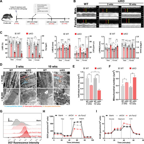
Defective myocardial and mitochondrial functions in Fars2 deficiency models. A, Schematic diagram of experimental protocol in mice. B, Representative M-mode echocardiographic images from the mice 3 and 10 weeks after inducible cardiac-specific Fars2 knockout (icKO). End-systole stages are indicated by red lines and end-diastole stages by yellow lines. C, Echocardiographic quantifications of wild-type (WT) and icKO mice. Shown in the statistical graph are left ventricular ejection fraction (LVEF) and left ventricular fractional shortening (LVFS; left panel), left ventricular systolic internal diameters (LVID;S) and left ventricular volume at end-systole (LV vol;S; middle panel), and left ventricular posterior wall thickness at end-diastole (LVPW;D) and left ventricular posterior wall thickness at end-systole (LVPW;S; right panel). D, Mitochondrial and myocardial dysmorphometry evoked by FARS2 (mitochondrial phenylalanyl-tRNA synthetase) deficiency. Transmission electron microscopy images (×15 000 and ×80 000); representative cardiomyocyte mitochondria are emphasized by white lines. A representative intermyofibrillar edema area is indicated by blue lines, a representative disarrayed myofilaments area by red lines, and a representative disrupted mitochondria area by orange lines. Top panel scale bar=2 μm; bottom panel scale bar=200 nm. E, Quantification of the average individual mitochondrial area (μm2) from D (n=3). F, Quantification of the average mitochondrial number (μm2) from D (n=3). G, Increase of reactive oxygen species production in Fars2 knockdown neonatal rat ventricular myocytes at 3, 5, and 7 days by DCFH-DA staining. H, Oxygen consumption rate in neonatal rat ventricular myocytes infected with shCtrl or sh-Fars2 at 3 days. I, Extracellular acidification rate in neonatal rat ventricular myocytes infected with shCtrl or sh-Fars2 at 3 days.

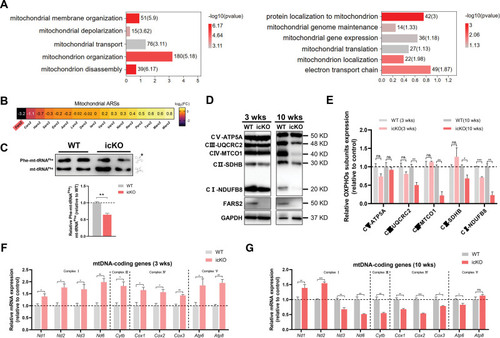
Defective mitochondrial homeostasis and protein synthesis in icKO hearts. A, Gene Ontology analysis of mitochondrial homeostasis–related biology process changes. B, Heatmap of mitochondrial aminoacyl-tRNA synthetase (mtARS) genes of inducible cardiac-specific Fars2 knockout (icKO) compared with wild-type (WT) mice from RNA sequencing analysis. C, Top, aminoacylation assay for mitochondrial mt-tRNAPhe in mouse heart. Charged (Phe-mt-tRNAPhe) and uncharged tRNA (mt-tRNAPhe) are indicated. Bottom, quantification of Phe-mt-tRNAPhe/mt-tRNAPhe from the top panel (n=3). D, Western blots of oxidative phosphorylation (OXPHOS) complexes and Fars2 in WT and icKO mice. E, Quantification of the OXPHOS complexes from D (n=3). F and G, Expression levels of mitochondrial DNA (mtDNA) coding genes in the heart of icKO mice at 3 weeks (F) and icKO mice at 10 weeks (G; n=3 mice per group). *P<0.05; **P<0.01; ***P<0.001.

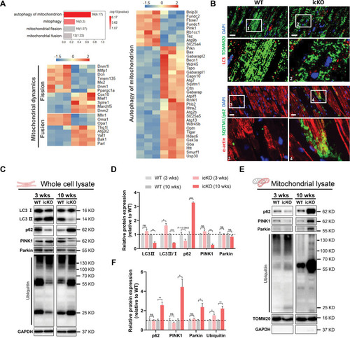
FARS2 deficiency causes MQC system disruption. A, Gene Ontology analysis of mitochondrial quality control (MQC) system–related biology process changes and heatmap of key genes in the autophagy of mitochondrion and mitochondrial dynamics pathways from RNA sequencing analysis. B, Top, Representative immunofluorescence images of LC3 (red) and TOMM20 (green) from mouse heart 10 weeks after inducible cardiac-specific Fars2 knockout (icKO) or in wild-type (WT) mice. Bottom, Representative immunofluorescence images of α-actin (red) and SQSTM1/p62 (green) from mouse heart 10 weeks after icKO or in WT mice. Scale bar=20 μm. C, Western blots of proteins in autophagy of mitochondria in whole-cell lysate of WT and icKO mice. D, Quantification of relative protein expression in C (n=3). E, Western blots of proteins in autophagy of mitochondria in mitochondrial lysate of WT and icKO mice. F, Quantification of relative protein expression in E (n=3). *P<0.05; **P<0.01; ***P<0.001.

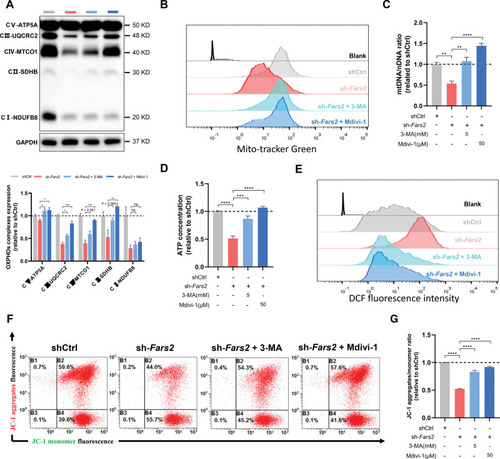
3-MA and Mdivi-1 attenuate mitochondrial dyshomeostasis induced by FARS2 deficiency in neonatal rat ventricular myocytes. A, Top, Western blots of oxidative phosphorylation (OXPHOS) complexes of control and Fars2 knockdown neonatal rat ventricular myocytes (NRVMs) after 3-methyladenine (3-MA) or Mdivi-1 treatment. Bottom, Quantification of the OXPHOS complexes from the top panel (n=3). B, Increased mitochondrial mass in Fars2 knockdown NRVMs after 3-MA or Mdivi-1 treatment by mitoTracker Green staining. C, Mitochondrial DNA copy number (mtDNA-CN; mtDNA/nuclear DNA) was detected by quantitative reverse transcription polymerase chain reaction of NRVMs after 3-MA or Mdivi-1 treatment (n=3). D, Quantification of the relative ATP contents of Fars2 knockdown NRVMs after 3-MA or Mdivi-1 treatment (n=3). E, Reactive oxygen species (ROS) production in Fars2 knockdown NRVMs at 3, 5, and 7 days after 3-MA or Mdivi-1 treatment by DCFH-DA staining. F, Representative FARS2 (mitochondrial phenylalanyl-tRNA synthetase) images in Fars2 knockdown NRVMs after 3-MA or Mdivi-1 treatment by JC-1 staining. G, Quantification of relative JC-1 aggregates/monomer ratio from F (n=3). *P<0.05; **P<0.01; ***P<0.001; ****P<0.0001.

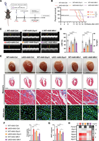
AAV9-mediated Drp1 knockdown or Mfn1 overexpression attenuates myocardial dysfunction induced by Fars2 deficiency and prolongs life span in mice. A, Schematic diagram of experimental protocol of adeno-associated virus 9 (AAV9)–mediated rescue tactics for inducible cardiac-specific Fars2 knockout (icKO) mice. B, Kaplan-Meier survival curves for wild-type (WT) and icKO mice after AAV9 treatment (n =9 per group). The median survival times for each group are marked. C, M-mode echocardiographic images from each group of mice at 10 weeks after icKO. End-systole stages are indicated by red lines and end-diastole stages by yellow lines. D, Left ventricular ejection fraction (LVEF; left panel) and left ventricular fractional shortening (LVFS; right panel; n=7–11 mice per group). E, Representative hearts (scale bar=2 mm): longitudinal sections (scale bar=2 mm), Masson trichrome staining (scale bar=25 μm), hematoxylin & eosin staining (scale bar=25 μm), and wheat germ agglutinin (WGA) staining (scale bar=25 μm) from each group 10 weeks after icKO. F, Quantification of average cardiomyocyte sectional size from E (at least 100 cells from 3 mice per group). G, Ratios of heart weight to tibia length (HW/TL; mg/mm) in different groups (n=6). H, Relative mRNA levels of Fars2, Drp1, Mfn1, and cardiac hypertrophy markers (Nppa, Nppb, and Myh7) in mice 10 weeks after icKO (n=3). *P<0.05; **P<0.01; ***P<0.001; ****P<0.0001.

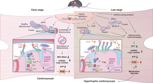
FARS2 deficiency causes cardiomyopathy by disrupting mitochondrial homeostasis and the mitochondrial quality control system. ETC indicates electron transport chain; FARS2, mitochondrial phenylalanyl-tRNA synthetase. mtDNA, mitochondrial DNA; and ROS, reactive oxygen species.

Unillustrated author statements

PHENOTYPE:
Fish:
Knockdown Reagents:
Observed In:
Stage: Long-pec
Acknowledgments
This image is the copyrighted work of the attributed author or publisher, and ZFIN has permission only to display this image to its users. Additional permissions should be obtained from the applicable author or publisher of the image. Full text @ Circulation