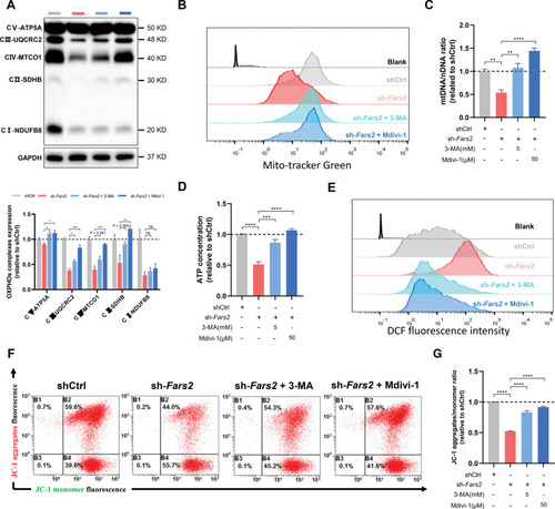
3-MA and Mdivi-1 attenuate mitochondrial dyshomeostasis induced by FARS2 deficiency in neonatal rat ventricular myocytes. A, Top, Western blots of oxidative phosphorylation (OXPHOS) complexes of control and Fars2 knockdown neonatal rat ventricular myocytes (NRVMs) after 3-methyladenine (3-MA) or Mdivi-1 treatment. Bottom, Quantification of the OXPHOS complexes from the top panel (n=3). B, Increased mitochondrial mass in Fars2 knockdown NRVMs after 3-MA or Mdivi-1 treatment by mitoTracker Green staining. C, Mitochondrial DNA copy number (mtDNA-CN; mtDNA/nuclear DNA) was detected by quantitative reverse transcription polymerase chain reaction of NRVMs after 3-MA or Mdivi-1 treatment (n=3). D, Quantification of the relative ATP contents of Fars2 knockdown NRVMs after 3-MA or Mdivi-1 treatment (n=3). E, Reactive oxygen species (ROS) production in Fars2 knockdown NRVMs at 3, 5, and 7 days after 3-MA or Mdivi-1 treatment by DCFH-DA staining. F, Representative FARS2 (mitochondrial phenylalanyl-tRNA synthetase) images in Fars2 knockdown NRVMs after 3-MA or Mdivi-1 treatment by JC-1 staining. G, Quantification of relative JC-1 aggregates/monomer ratio from F (n=3). *P<0.05; **P<0.01; ***P<0.001; ****P<0.0001.
|