FIGURE SUMMARY
Title

Translational control of furina by an RNA regulon is important for left-right patterning, heart morphogenesis and cardiac valve function

Authors
Nagorska, A., Zaucker, A., Lambert, F., Inman, A., Toral-Perez, S., Gorodkin, J., Wan, Y., Smutny, M., Sampath, K.
Source
Full text @ Development

Zebrafish furina transcripts show different 3′UTR lengths, with a predominant long isoform expressed during somitogenesis. (A) Schematic of furina transcript 1 and variant X1. Orange arrowheads represent primers to amplify transcript 1; one orange and one green primer were used to detect variant X1. Hairpin indicates the YBE motif in the 3′UTR of X1. (B) RNA sequencing excerpt from ovary, 50% epiboly and 14-19 som stages. (C) Semi-quantitative RT-PCR detecting gapdh positive control, furina coding sequences (CDS) and furina variant X1 3′UTR (X1). (D) Schematic of primers used in qPCR to detect the furina CDS and variant X1. The orange primer set was used to amplify the CDS and green primers used to amplify variant X1. (E) Quantitative PCR analysis from ovary, 1 K, 50% epiboly, 10-som and 18-som embryos. Orange bars represent furina coding region, green bars show exclusively variant X1. **P<0.01 in unpaired, two-tailed Student's t-test. n=50 embryos per stage for mRNA extraction, P=0.014. ns, not significant. (F) Whole-mount in situ hybridisation with probes that detect the CDS or variant X1. Yellow arrowheads point to enrichment in eyes, red in hindbrain and blue in somites. Lateral and dorsal views are shown at 10-som and 18-som. Scale bars: 100 µm.

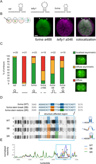
The furina variant X1 transcript harbours a YBE motif in the 3′UTR. (A) schematic showing hairpin YBE motifs in sqt, lefty1 and furina variant X1. (B) Left: schematic of localisation assay. Embryos were injected into the yolk at the 1-cell stage and mRNA localisation was analysed at the 4-cell stage. Right: confocal images showing colocalisation of furina (labelled with Alexa 488-UTP) fluorescent reporter and lefty1 (labelled with Alexa 546-UTP) in zebrafish embryos at the 4-cell stage. n=11. Arrowheads indicate fluorescent reporter mRNA localisation. Scale bar: 20 µm. (C) Stacked bar graph showing localisation categories in fluorescent reporter mRNA-injected embryos: localised asymmetric (green), diffuse asymmetric (yellow) and diffuse (red). Schematic representations of fluorescent RNAs of control sqt (n=20), lacZ (n=21) and furina variant X1 RNAs (full-length furina; n=23), or disrupting the YBE motif (ΔYBE; n=19; SB, n=22; SR, n=19) are shown below in black. (D) Structure probing of furina X1 3′UTR. Structural mutations were generated based on the predicted stem loop (SB, SR). Structure footprint gels for furina RNAs. Blue highlighted sequences (top) and blue dotted box (bottom) indicates the structure affected region. Lanes labelled C indicate control untreated RNA that was reverse transcribed; lanes labelled T indicate RNA treated with NAI-N3 before reverse transcription. Dashed lines separate cropped regions from different parts of the same gel. Schematics on the right represent the predicted YBE structure in WT, SB and SR. (E) Quantification of structure footprint gels. Blue dotted box represents the structure affected region (blue highlighted sequences). Black bars indicate the predicted YBE stem loop structure.

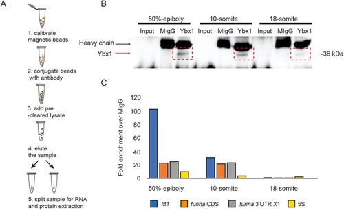
Ybx1 protein interacts with furina variant X1 mRNA. (A) Schematic of RNA immunoprecipitation of embryo lysates with anti-Ybx1 antibodies. (B) Representative western blot showing pull-down of Ybx1 from 50% epiboly, 10-som and 18-som embryonic lysates. n=250 embryos per stage; black arrow indicates IgG heavy chain; red arrow indicates Ybx1 band. (C) qPCR analysis of samples from immunoprecipitation. Fold enrichment normalised to input and mIgG control are shown for lefty1 (blue), furina coding region (orange), furina variant X1 3′UTR (grey) and 5S negative control (yellow).

FurinA translation and Spaw maturation is increased in ybx1 mutant embryos. (A) Schematic of GFP fusion reporters for furina X1 and spaw. Hairpin indicates the YBE motif in furina RNA. The FurinA pro-domain is shown in dark grey and the signal peptide in yellow; for Spaw, the pro-domain is indicated in dark grey; stop codon indicated in red in FurinA and Spaw. (B) Schematic of temperature shift of ybx1sa42 mutant embryos. Embryos were injected with GFP reporter-fusion mRNA, grown at 28°C until the 16-cell stage, shifted to 22°C until the 512-cell stage, when protein was extracted. (C) Western blots of embryonic lysates showing FurinA-sfGFP (left) and Spaw-GFP fusion protein (right), following injection of embryos with mRNA reporter. Lanes were loaded with lysates from un-injected wild-type control, Mybx1 at 22°C and Mybx1 control embryos at 28°C. Actin loading control is aligned to the corresponding samples. (D) Bar graphs show quantification of corresponding band intensities, with FurinA-sfGFP (left) and mature Spaw (right) normalised to the Actin loading control, *P<0.05 (P=0.021 for FurinA-GFP; P=0.026 for Spaw-GFP). (E) Schematic showing temperature shift of ybx1 mutant embryos following injection with spaw GFP reporter. (F) Confocal images of 1 K, sphere and 30% epiboly wild-type (top) and ybx1 mutant (bottom) embryos. WT, wild type. Scale bars: 50 µm.

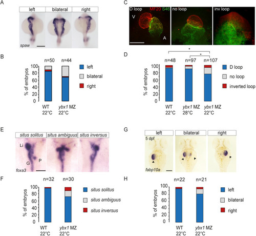
Mutant ybx1sa42 embryos have abnormal LR expression. (A) WISH to detect spaw in wild-type (n=50) and ybx1sa42 mutant (n=44) embryos at the 21-som stage. Scale bar: 100 µm. (B) Bar graph showing the proportion of embryos with spaw expression on the left (blue), bilaterally (grey) or on the right (red). Fisher's Exact two-tailed probability test P=0.0125. (C) Immunofluorescence showing MF20 labelling in the ventricle (red) and S46 labelling in the atrium (green) at 55 hpf. Representative D loop, no loop and inverted (inv) loop hearts are shown. A, atrium; V, ventricle. Scale bar: 100 µm. (D) Quantification of looping of the heart. Directional loop (D loop, blue), no loop (grey) and inverted looping (red) of the heart, with ybx1 mutant embryos at 22°C (n=107) showing a higher percentage of inverted and no loop hearts compared with control wild-type (WT; n=48) or ybx1 embryos at 28°C (n=97). An unpaired, two-tailed Student's t-test was used to analyse the data. *P<0.05. MZybx1 22°C compared with 28°C: P=0.022; MZybx1 22°C compared with WT: P=0.030. (E) WISH to detect foxa3 at 55 hpf in wild-type and ybx1 mutant embryos. G, gut; Li, liver; P, pancreas. Scale bar: 100 µm. (F) Quantification of visceral organ positioning in wild-type (n=32) and ybx1 mutant (n=30) embryos at 55 hpf showing an increase in situs ambiguus (grey) and situs inversus (red) with a decrease in situs solitus (blue) in ybx1 mutant embryos. Fisher's Exact two-tailed test. P=0.0113. (G) Expression of fabp10a at 5 dpf. Arrowheads indicate the liver. Scale bar: 100 µm. (H) Quantification of liver positioning in wild-type (n=22) and ybx1 mutant (n=21) embryos showing situs solitus (blue), situs ambiguus (grey) and situs inversus (red).

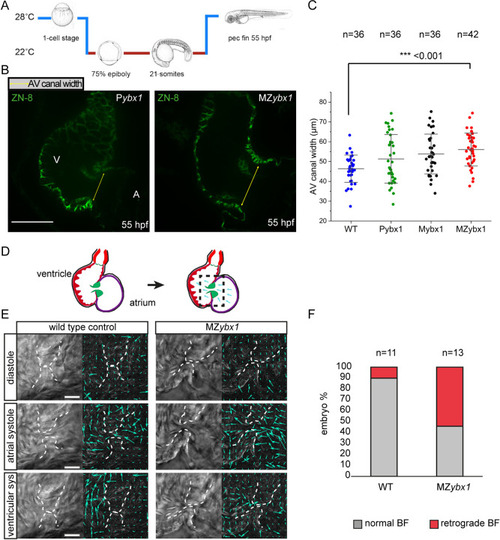
Ybx1 mutant embryos show abnormal heart morphogenesis. (A) Schematic of temperature shift experiments. ybx1 mutant embryos and controls were shifted to 22°C at 75% epiboly until 21-som and then grown at 28°C until 55 hpf. (B) Confocal images of zebrafish heart showing immunofluorescence with the ZN-8 antibody to label the of atrioventricular (AV) canal. Yellow arrows indicate the width of the AV canal in control Pybx1 and MZybx1 mutant embryos. A, atrium; V, ventricle. Scale bar: 25 µm. (C) Quantification AV canal width in µm from immunostaining of wild-type (WT; blue, n=36), Pybx1 (green, n=36), Mybx1 (black, n=36) and MZybx1 embryos (red, n=42). ANOVA test was used for statistical analysis (P=0.00016361; ***P<0.001). (D) Schematic of zebrafish heart at 5 dpf (left) and schematic of PIV analysis (right); black dashed box shows the area of the heart imaged and analysed. (E) Left: DIC images from movies of wild-type and ybx1 mutant hearts during diastole, atrial systole and ventricular systole. Right: PIV analysis of the images; cyan arrows indicate movements of red blood cells. Scale bars: 20 µm. (F) Percentage of embryos showing normal blood flow (BF) versus retrograde BF in wild-type (n=11) and ybx1 mutant (n=12) embryo groups.

Mutant ybx1 embryos have retrograde blood flow at 5 dpf. (A) Schematic of a zebrafish heart at 5 dpf. Arrows indicate the directions of flow; black box shows the region analysed in PIV. (B) Representative image of a wild-type (WT) embryo, showing PIV application at 0.038 s of ventricular systole. Vectors generated from red blood cells are shown as blue arrows. Rose plot shows quantification of direction of the blood flow at 0.038 s for the imaged wild-type hearts (n=11). Scale bar: 25 µm. (C) A PIV image of wild-type ventricular contraction at 0.173 s (left) and quantification of direction of generated vectors (rose plot on right) (n=11). Vectors are shown as blue arrows. Scale bar: 25 µm (bars in B,D apply to C,E, respectively). (D) A PIV image showing the direction of blood flow in MZybx1 embryos with retrograde blood flow at 0.038 s. Vectors are shown as red arrows. Rose plot shows analysis of direction of blood flow in MZybx1 embryos with retrograde blood flow (n=6). Scale bar: 25 µm. (E) PIV analysis at 0.173 s of ventricular systole and quantification of vector direction in ybx1 embryos with retrograde blood flow at 0.173 s (n=6). Vectors are shown as red arrows. Scale bar: 25 µm. Dashed lines in B-E delineate heart valves. (F) Analysis of average velocity of horizontal vectors moving towards the atrium over the duration of ventricular contraction. Wild-type embryos are shown in blue and ybx1 mutants in red. (G) Analysis of average velocity of vertical vectors moving out of the ventricle during ventricular contraction. Wild-type embryos in blue and ybx1 mutants in red.

Deletion of the furina 3′UTR YBE leads to abnormal LR patterning. (A) Schematic of the furina X1 3′UTR genomic region on chromosome 7. Exon 16 contains the 3′ end of the CDS with the stop codon (stop sign) and the entire 3375 bp of the 3′UTR. The red dotted line shows the sequences deleted in furina Δ3′UTR crispants by the two gRNAs (green arrows). The position of primers used for the simultaneous detection of 3′UTR deletion alleles (primer F and primer 2R) with wild-type alleles (primer F and primer 1R) in three primer genotyping PCRs are indicated. (B) Gel image showing furina Δ3′UTR crispant genotyping PCRs using genomic DNA from 1 dpf embryos. The expected sizes of the wild-type (wt) and the Δ3′UTR deletion mutant PCR products are shown next to the 100 bp DNA ladder (far-left lane). Mutant bands (cyan arrowheads) together with wild-type bands are only observed with the crispant samples. The non-injected control (Nij) only shows the wild-type band, and no product is detected in the no template control lane (NTC). (C) Stacked bar chart showing the percentage of embryos falling into different categories for spaw expression by WISH on 19-21 som embryos: non-injected wild-type embryos, Cas9 protein only-injected embryos, Cas9 RNPs loaded with the two targeting gRNAs (gRNAs 1+2), and control Cas9 RNPs loaded with gRNA2 and a non-cutting control gRNA (gRNAc). n=number of embryos. *P<0.05 by Fisher's exact test. (D) Images of spaw expression by WISH in 19-21 som embryos showing the various categories shown in the bar graph in C.

Acknowledgments
This image is the copyrighted work of the attributed author or publisher, and ZFIN has permission only to display this image to its users. Additional permissions should be obtained from the applicable author or publisher of the image. Full text @ Development