IMAGE

Fig. 3.

ID
ZDB-IMAGE-231220-63
Source
Figures for Nagorska et al., 2023
Image
Figure Caption

Fig. 3.

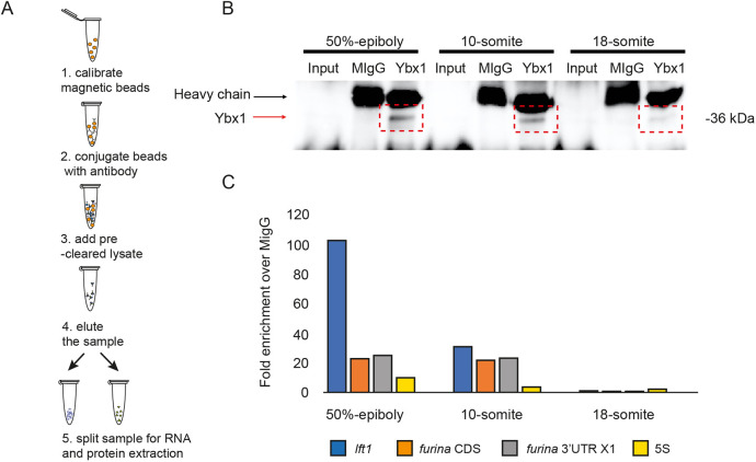
Ybx1 protein interacts with furina variant X1 mRNA. (A) Schematic of RNA immunoprecipitation of embryo lysates with anti-Ybx1 antibodies. (B) Representative western blot showing pull-down of Ybx1 from 50% epiboly, 10-som and 18-som embryonic lysates. n=250 embryos per stage; black arrow indicates IgG heavy chain; red arrow indicates Ybx1 band. (C) qPCR analysis of samples from immunoprecipitation. Fold enrichment normalised to input and mIgG control are shown for lefty1 (blue), furina coding region (orange), furina variant X1 3′UTR (grey) and 5S negative control (yellow).

Acknowledgments
This image is the copyrighted work of the attributed author or publisher, and ZFIN has permission only to display this image to its users. Additional permissions should be obtained from the applicable author or publisher of the image. Full text @ Development