- Title
-
p53-Dependent DNA damage response sensitive to editing-defective tRNA synthetase in zebrafish
- Authors
- Song, Y., Shi, Y., Carland, T.M., Lian, S., Sasaki, T., Schork, N.J., Head, S.R., Kishi, S., Schimmel, P.
- Source
- Full text @ Proc. Natl. Acad. Sci. USA
|
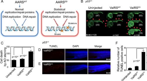
ED ValRS causes cell death and DNA damage in zebrafish. (A) The ED tRNA synthetase is proposed to generate statistical replication and repair proteins that lead to increased DNA breaks. (B) AO staining at 1 dpf of zebrafish uninjected (n = 120), or injected with either ValRSWT mRNA (n = 142) or ValRSED mRNA (n = 136). The red arrows show points of cell death. Zebrafish are aligned vertically based on cell death severity and morphology; normal (Top), increased cell death (Middle), and increased cell death with abnormal morphology (Bottom). (Scale bar, 500 µm.) (C) Quantification of AO staining to determine average number of cell deaths per fish. Bars represent mean ± SEM. Images of TUNEL staining on fish injected with either (D) ValRSWT or (E) ValRSED mRNA. TUNEL staining, DAPI staining, and TUNEL merged with DAPI, are shown. (Scale bars, 100 µm.) (F) Quantification of the average number of TUNEL+ cells per fish (periderm or basal epidermal cells on the trunk). Independent areas [from the trunk region starting at the rostral start point of the yolk extension (the distal end of yolk) and extending through the end of the caudal fin] were selected from individual animals that had been injected with mRNA and were used for the quantification. Quantifications (Uninjected n = 13, ValRSWT n = 16, ValRSED n = 23) are shown in the graph. Bars represent mean ± SEM. |
|
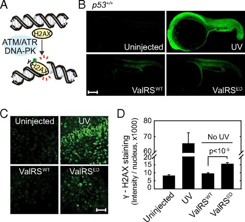
Response of H2AX to an ED ValRS. (A) Cartoon of H2AX phosphorylation. (B) γ-H2AX staining in uninjected (n = 60), ValRSWT mRNA injected (n = 65), ValRSED mRNA injected (n = 79), or UV treated zebrafish (n = 30) at 1 dpf. (Scale bar, 250 µm.) (C) Enlarged pictures of γ-H2AX staining in the nuclei of different samples of zebrafish. (Scale bar, 40 µm.) (D) Quantification of γ-H2AX staining. Bars represent mean ± SEM. |
|
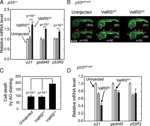
ED ValRS activates p53 in zebrafish. (A) RT-PCR results of p53 downstream markers p21, gadd45, and p53R2 in ValRSWT or ValRSED mRNA-injected zebrafish. The bar graphs show the mean values ± SEM after normalization to the β-actin level. (B) Uninjected (n = 118), ValRSWT mRNA-injected (n = 90), or ValRSED mRNA-injected (n = 93) p53zdf1/zdf1 zebrafish were stained with AO at 1 dpf. The red arrows show points of cell death. Zebrafish are aligned based on cell death severity. (Scale bar, 500 µm.) (C) Quantification of AO staining. Bars represent mean ± SEM. (D) RT-PCR results of p53 downstream markers p21, gadd45, and p53R2 in p53zdf1/zdf1 zebrafish injected with either ValRSWT or ValRSED mRNA. The bar graphs show the mean values ± SEM after normalization to the β-actin level. EXPRESSION / LABELING:
|
|
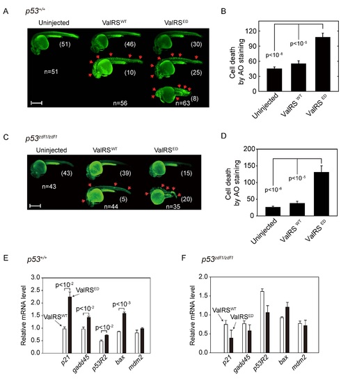
mValRSED mRNA injection into zebrafish results in cell death and p53 activation. A) Acridine orange (AO) staining at 1 dpf of zebrafish uninjected (n=51), or injected with either mValRSWT mRNA (n=56) or mValRSED mRNA (n=63). The red arrows show points of cell death. Zebrafish are aligned vertically based on cell death severity and morphology; normal (top), increased cell death (middle), and increased cell death with abnormal morphology (bottom). Scale bar represents 500 µm. B) Quantification of acridine orange staining per fish. Bars represent mean ± SEM. C) Uninjected (n=43), mValRSWT mRNA injected (n=44), or mValRSED mRNA injected (n=35) p53zdf1/zdf1 zebrafish were stained with acridine orange at 1 dpf. The red arrows show points of cell death. Zebrafish are aligned based on cell death severity. Scale bar represents 500 µm. D) Quantification of acridine orange staining. Bars represent mean ± SEM. RT-PCR results of p53 downstream markers p21, gadd45, p53R2, bax and mdm2 in E) p53+/+ or F) p53zdf1/zdf1 zebrafish injected with either mValRSWT or mValRSED mRNA. |
|
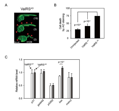
The effects of aminoacylation-deficient mValRS (ValRSAD) in zebrafish. A) Zebrafish injected with mValRSAD mRNA (n=24) were stained with acridine orange at 1 dpf. Zebrafish are aligned based on cell death severity and zebrafish morphology; normal (top), increased cell death (middle), increased cell death with abnormal morphology (bottom). Scale bar represents 500 µm. B) Quantification of acridine orange staining. Zebrafish were uninjected, or injected with mValRSWT or mValRSAD mRNAs. Bars represent mean ± SEM. C) RT-PCR results of p53 downstream markers p21, gadd45, p53R2, bax and mdm2 in zebrafish injected with mValRSWT or mValRSAD mRNA. All data are normalized to a value of 1.0 for the uninjected sample. |
|
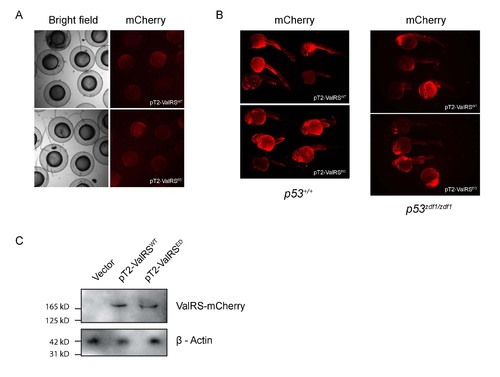
ValRS-mcherry expression test. A) Zebrafish injected with pT2-ValRSWT or ValRSED (both mcherry tagged) were collected at 8 hpf or B) 24 hpf and imaged for ValRS-mcherry expression. C) At 2 dpf zebrafish embryos injected with pT2-ValRSWT or ValRSED (both mcherry tagged) were lysed and ran on a SDS-PAGE gel, and subjected to western blot analysis with a mcherry antibody. |

