- Title
-
BMP signaling orchestrates photoreceptor specification in the zebrafish pineal gland in collaboration with Notch
- Authors
- Quillien, A., Blanco-Sanchez, B., Halluin, C., Moore, J.C., Lawson, N.D., Blader, P., and Cau, E.
- Source
- Full text @ Development
|
BMP activity is necessary for PhRs specification. (A,B) Confocal sections of control (A) and Tg(hs:dnBmpr1a-CFP);Tg(hsp70:noggin3) double transgenic (B) embryo. Pineal glands are double labeled with the Tg(AANAT2:GFP) transgene (in red) and a HuC/D antibody (in green) at 48 hours. Anterior is upwards. Scale bar: 16 μm. (C-E) Average numbers of Isl1+ neurons, Tg(AANAT2:GFP)+ and Tg(HuC:GFP)+ cells per pineal gland in control, Tg(hs:dnBmpr1a-CFP) transgenic, Tg(hsp70:noggin3) transgenic and Tg(hs:dnBmpr1a-CFP);Tg(hsp70:noggin3) double-transgenic embryos at 48 hours. Error bars represent s.d. **P<0.001; ***P<0.0005 using a t-test. The number (n) of embryos analyzed is noted for each case. (F-G′′) Confocal images of the pineal gland of wild-type host embryos that have received cells transplanted from wild type (F-F′′) or Tg(hs:dnBmpr1a-CFP) (G-G′′) donors. Tg(AANAT2:GFP)+ PhRs are in green and the transplanted cells are shown in red. White arrowheads show transplanted cells with a PhR identity; white arrows indicate transplanted cells not expressing GFP from the Tg(AANAT2:GFP) transgene. EXPRESSION / LABELING:
|
|
Bmp2a is required for PhR specification. (A-D) Expression of bmp2a in the wild-type pineal at 14, 16, 18 and 20 hours. (E-H′) Activation of the BMP pathway as judged by labeling with an α-PSmad 1/5/8 antibody shown on confocal projections. The presumptive pineal territory is delineated by GFP expression from the Tg(flh:gfp) transgenic line (Concha et al., 2003). Anterior is upwards. Scale bars: 16 μm. (I-O) Average numbers of Isl1+ neurons, Tg(AANAT2:GFP)+, FRet43+, HuC/D+, Tg(HuC:GFP)+, Isl1+/Pax6+ and lhx3+ cells per pineal gland at 48 hours in control, bmp2a and smad5 morpholino-injected embryos. The number (n) of embryos analyzed is noted for each case. Error bars represent s.d. **P<0.001, ***P<0.0005 using a t-test. EXPRESSION / LABELING:
PHENOTYPE:
|
|
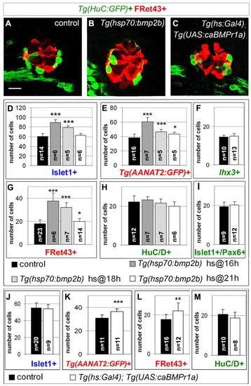
BMP activity is sufficient to promote the PhR fate. (A-C) Confocal sections of control, Tg(hsp70:bmp2b) transgenic and Tg(hs:Gal4);Tg(UAS:caBmpr1a) double transgenic embryos at 48 hours. Pineal glands are double-stained for Tg(HuC:GFP) (in green) and FRet43 (in red). Anterior is upwards. Scale bar: 16 μm. (D-I) Average numbers of Isl1+ neurons, Tg(AANAT2:GFP)+, lhx3+, FRet43+, HuC/D+ and Isl1+/Pax6+ cells in the pineal gland of control and transgenic Tg(hsp70:bmp2b) embryos heat-shocked at various stages and analyzed at 48 hours. In E and G, Tg(hs:bmp2b) embryos heat-shocked at 21 hours show a relatively mild but statistically significant effect (P=0.03 and P=0.037, respectively). (J-M) Average numbers of Isl1+ neurons, Tg(AANAT2:GFP)+, Fret43+ and HuC/D+ cells in the pineal gland of control and Tg(hs:Gal4); Tg(UAS:caBmpr1a) double-transgenic embryos at 48 hours. Heat shocks were performed at 16 hours. Error bars represent s.d. *P<0.05, **P<0.001, ***P<0.0005 using a t-test. EXPRESSION / LABELING:
|
|
BMP activity is necessary and sufficient for the expression of regulators of the PhR fate. (A-D′) Confocal sections of pineal glands, showing expression of neurod1 or otx5 (in red) and GFP (in green) from Tg(AANAT2:GFP) (A,A′,C,C′) and Tg(HuC:GFP) (B,B′,D,D′) transgenic embryos at 24 hours. White arrowheads indicate double-labeled cells. neurod1 and Tg(AANAT2:GFP) are co-expressed in the majority of cells, although single labeled cells are also present. The observation of neurod1+/Tg(AANAT2:GFP)- cells most probably reflects the earlier onset of expression for neurod1 compared with that of the Tg(AANAT2:GFP) transgene (Gothilf et al., 2002). Conversely, neurod1-/Tg(AANAT2:GFP)+ could be PhRs that do not express neurod1 during their life or alternatively more mature cells that have already turned off the gene. Similarly, although all Tg(AANAT2:GFP)+ cells are also otx5 positive, a number of single-labeled otx5+ cells are observed, which is probably due to the early onset of otx5 (see below) compared with the Tg(AANAT2:GFP) transgene. (E-L) Dorsal view of pineal glands stained for neurod1 (E-H) or otx5 (I-L) by in situ hybridization. Both genes start to be expressed at 16 hours. (M-O) Dorsal view of pineal gland from wild-type (M), Tg(hs:dnBmpr1a-CFP); Tg(hsp70:noggin3) double transgenic (N) and Tg(hs:bmp2b) embryos (O) at 24 hours stained for neurod1. (P) Quantification of the data represented in M-O. (Q-S) Dorsal view of pineal glands from wild-type (Q), Tg(hs:dnBmpr1a-CFP); Tg(hsp70:noggin3) double transgenic (R) and Tg(hs:bmp2b) embryos (S) at 24 hours stained for otx5. (T) Quantification of the data represented in Q-S. In M-T, Tg(hs:dnBmpr1a-CFP); Tg(hsp70:noggin3) transgenics were heat shocked at 16 hours, while Tg(hs:bmp2b) were heat shocked at 21 hours. The number (n) of embryos analyzed is noted for each case in P and T. Error bars represent s.d. ***P<0.0005 using a t-test. Anterior is upwards. Scale bars: 16 μm. EXPRESSION / LABELING:
|
|
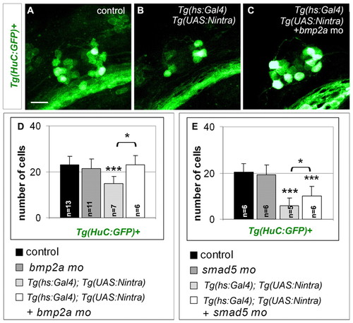
BMP regulates the competence to respond to Notch. (A-C) Confocal projections of control (A), Tg(hs:Gal4);Tg(UAS:Notchintra) transgenic (B) and Tg(hs:Gal4);Tg(UAS:Notchintra) transgenic bmp2a-morphant (C) embryos at 48 hours. Heat shock was performed at 14 hours. The effects of the various treatments were monitored by GFP immunostaining in the Tg(HuC:GFP) background. (D,E) Average numbers of Tg(HuC:GFP)+ cells in 48 hour control, Tg(hs:Gal4);Tg(UAS:Notchintra) transgenic, Tg(hs:Gal4);Tg(UAS:Notchintra) transgenic bmp2a-morphant and bmp2a-morphant (D) embryos or smad5-morphant embryos (E). Heat shocks were performed at 14 hours. The number (n) of embryos analyzed is noted for each case in D and E. Error bars represent s.d. *P<0.05, ***P<0.0005 using a t-test. Anterior is upwards. Scale bar: 16 μm. EXPRESSION / LABELING:
|
|
BMP regulates Notch target gene expression. (A-I) Dorsal view of pineal glands showing expression of her genes in control (A,D,G), mib (B,E,H) and Tg(hs:dnBmpr1a-CFP);Tg(hsp70:noggin3) transgenic embryos (C,F,I) at 20 hours. For Tg(hs:dnBmpr1a-CFP);Tg(hsp70:noggin3) transgenic embryos, heat shocks were performed at 16 hours. (J-L) Dorsal view of pineal glands from mock-treated (J), DAPT-treated (K) or smad5-morphant (L) embryos. The effects of the various treatments were monitored by in situ hybridization against gfp transcripts in a Tg(Tp1bglob:eGFP) background. Anterior is upwards. Scale bar: 16 μm. (M-N′) Confocal sections of pineal glands of wild-type host having received cells transplanted from wild-type (M-M′) or Tg(hs:dnBmpr1a-CFP) donors (N-N′). Transplanted cells are shown in green, while expression of her4 mRNA is shown in red. White arrowheads highlight transplanted cells expressing her4. EXPRESSION / LABELING:
|
|
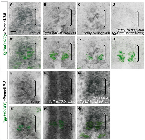
Levels of BMP activity upon induction of the various transgenes. Confocal projections from control (A,A′,E,E′), Tg(hs:dnBMPr1a-CFP) transgenic (B,B′), Tg(hsp70:noggin3) transgenic (C,C′), Tg(hs:dnBMPr1a-CFP);Tg(hsp70:noggin3) double-transgenic (D,D′); Tg(hs:BMP2b) transgenic (F,F′) and Tg(hs:Gal4); Tg(UAS:caBMPr1a) double-transgenic (G-G′) embryos double-labeled with an α-PSmad 1/5/8 antibody (grey) and a Tg(HuC:GFP) transgene at 20 (A-D′) and 24 hours (E-G′). The Tg(HuC:GFP) transgene helps delineating the posterior limit of the pineal gland. The pineal area is indicated with a bracket. Transgenic and control embryos were heat shocked at 16 (A-D′) or 21 hours (E-G′). Anterior is upwards. Scale bars: 16 µm. |


