- Title
-
Chondroitin sulfate expression is required for cardiac atrioventricular canal formation
- Authors
- Peal, D.S., Burns, C.G., MacRae, C.A., and Milan, D.
- Source
- Full text @ Dev. Dyn.
|
Whole mount immunofluorescence images of Danio rerio hearts stained with an anti-chondroitin sulfate antibody at the indicated hours post fertilization (hpf). |
|
Confocal images of Danio rerio hearts stained with an anti-chondroitin sulfate antibody at 36 and 48 hours postfertilization (hpf). A,B: Confocal microscopy of Danio rerio cis/trans-decahydro-2-napthol-β-D-xyloside (DX) containing the myocardium specific cmlc2:GFP marker (green). C,D: Confocal microscopy of the same Danio rerio images stained with a CS-specific antibody (red). E,F: Merged images. a, atrium; v, ventricle; avc, atrioventricular canal forming region; oft, outflow tract. |
|
Chondroitin sulfate (CS) is localized to the cardiac jelly between the myocardium and endocardium. A: Confocal microscopy of showing a circumferential cross-section of 36 hours postfertilization (hpf) Danio rerio containing the myocardium specific cmlc2:GFP marker (green) and stained with a CS-specific antibody (red). B: Confocal microscope image of Danio rerio containing the endocardium specific flk:EGFP marker (green) and stained with a CS-specific antibody (red). |
|
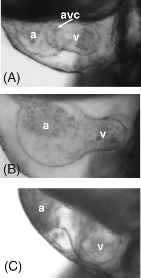
Chemical and genetic knockdown of chondroitin sulfate prevents the proper formation of the atrioventricular (AV) canal in zebrafish. A: Wild-type Danio rerio heart at 55 hours postfertilization (hpf). B: The 55 hpf animals treated from 7-48 hpf with DX (cis/trans-decahydro-2-napthol-β-D-xyloside), an inhibitor of CS formation. C: 55 hpf animals injected with a morpholino to chondroitin synthase-1 (chys-1). at. atrium; v, ventricle; av, atrioventricular canal forming region. PHENOTYPE:
|
|
Depletion of chondroitin sulfate leads to changes in the cell migration marker spp1. A: In situ hybridization section demonstrating that the zebrafish osteopontin marker spp1 is localized to the atrioventricular canal endocardium in zebrafish. B: spp1 is normally expressed in the atrioventricular canal (avc) and outflow tract (oft). C: DX (cis/trans-decahydro-2-napthol-β-D-xyloside) treatment from 7 to 48 hours postfertilization (hpf) results in the disappearance of spp1 in the AVC and OFT at 72 hpf. Note that, while spp1 expression is absent in DX treated embryos, the presence of a stripe of spp1 expression at the base of the pectoral fin demonstrating the success of the in situ protocol (white arrow in inset of B and C). EXPRESSION / LABELING:
|
|
Depletion of chondroitin sulfate results in disruption of multiple signaling pathways. A,C,E: Tbx2b, versican, and notch1b are normally expressed within the atrioventricular canal (AVC) at 48 hours postfertilization (hpf; black arrows). B,D,F: However, treatment with the compound DX results in the severe down-regulation of expression. G,H: Bmp4 expression remains within the AVC region when treated with DX (black arrows in G,H), but ventricular expression is up-regulated (white arrow in H). For those genes that are down-regulated, we have included an image of the entire upper body, as all three of these genes stain the head. This is to demonstrate the absence of heart expression is not due to failure of the in situ probe. EXPRESSION / LABELING:
|
|
Expression of chondroitin sulfate in whole mount zebrafish. Animals were stained with an antibody reactive to chondroitin sulfate. Dynamic changes in body expression patterns include early expression within the developing cardiovascular system (vas) and head, followed by later expression within the lateral line neuromasts (n), the pectoral fins (p), otic placodes (0), and pharyngeal arches. EXPRESSION / LABELING:
|
|
Immunfluorescence staining using the anti-myosin antibody S46 serves as a positive control for antibody staining at the 55 hpf time point. |
|
Chemical and genetic downregulation of chondroitin sulfate. (A) Confocal slice through the heart tube of wild type 36 hpf Danlo rerlo transgenic for the myocardial promoter cm/c2::GFP construct (green), counterstained with an antibody to chondroitin sulfate (red). (B) Animals treated with the CS-depleting compound DX show a reduction in cardiac CS. (C) Animals injected with a morpholino to chys-1 show an absence of cardiac CS. |
|
DX treatment does not deplete heparan sulfate (HS). (A) 36 hpf fish, DX treated (left) and non-treated (right), stained with an anti-HS antibody. The staining patterns and staining levels appear similar. (B) In order to demonstrate that this is not background staining, we compared jekyll mutants (left), which lack HS, to wild type (right). |

Unillustrated author statements |


