- Title
-
Genetic and cellular analyses of zebrafish atrioventricular cushion and valve development
- Authors
- Beis, D., Bartman, T., Jin, S.W., Scott, I.C., D'Amico, L.A., Ober, E.A., Verkade, H., Frantsve, J., Field, H.A., Wehman, A., Baier, H., Tallafuss, A., Bally-Cuif, L., Chen, J.N., Stainier, D.Y., and Jungblut, B.
- Source
- Full text @ Development
|
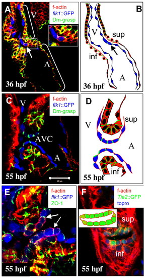
Cellular differentiation of wild-type AV canal between 36 and 55 hpf. Confocal images of the heart at 36 hpf (A) and the AV canal at 55 hpf (C,E,F). (A,C,E) Tg(flk1:EGFP)s843 (pseudo-colored blue) embryos stained with rhodamine phalloidin (red) and immunostained for Dm-grasp (pseudo-colored green) (A,C) or for ZO-1 (pseudo-colored green) (E). (F) Tg(Tie2:EGFP)s849 (green) embryo stained with topro (blue) and rhodamine phalloidin (red). (A) At 36 hpf, the heart has looped and the endocardium (in blue) is single layered and squamous. Arrow indicates one endocardial cell expressing Dm-grasp. (B) Schematic representation of the heart shown in A. (C) At 55 hpf, the AV canal endocardial cells exhibit a cuboidal shape and Dm-grasp is localized laterally. Ventricular and atrial endocardial cells appear squamous and devoid of Dm-grasp expression. Myocardial cells in the superior (sup) and inferior (inf) ECFR of the AV canal exhibit stronger expression of laterally localized Dm-grasp compared with ventricular and atrial myocardial cells. (D) Schematic representation of the AV canal shown in C. (E) ZO-1 is expressed by all myocardial and endocardial cells, including the cuboidal endocardial cells lining the AV canal. Arrows indicate ZO-1 localized in tight junctions between two neighboring cuboidal endocardial cells. (F) Transverse section. A five or six cell-wide sheet of cuboidal cells line the superior and inferior regions of the AV canal. Laterally and in this plane, two squamous cells (hinge cells; light green) connect the sheets of cuboidal cells. The inset is a schematic representation of the pattern of endocardial cell shapes across the AV canal. A, atrium; V, ventricle; AVC, atrioventricular canal; inf, inferior ECFR; sup: superior ECFR.
|
|
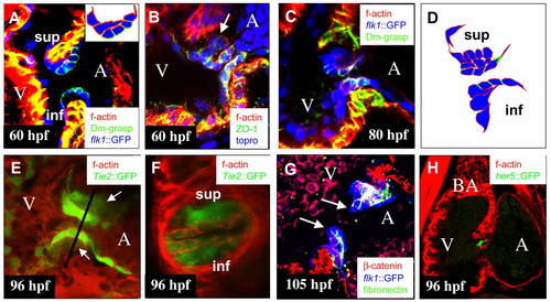
Wild-type endocardial cushion morphogenesis. Confocal images of the AV canal at 60 (A,B), 80 (C), 96 (E,F) and 105 hpf (G), and of the heart at 96 hpf (H). (A,C,G) Tg(flk1:EGFP)s843 (pseudo-colored blue) embryo immunostained for Dm-grasp (pseudo-colored green) and stained with rhodamine phalloidin (red) (A,C) or immunostained for fibronectin (pseudo-colored green) and ß catenin (red) (G). (E,F) Tg(Tie2:EGFP)s849 (green) embryo stained with rhodamine phalloidin (red). (B) Embryo immunostained for ZO-1 (green) and stained with topro (blue) and rhodamine phalloidin (red). (H) Tg(0.7her5:EGFP)ne2067 (green) embryo stained with rhodamine phalloidin (red). (A) AV endocardial cell at the ventricular border has extended into the ECM and is reaching the base of cells close to the atrial border. Inset shows schematic drawing of AV endocardial cells of the superior AV EC. (B) Cells, such as the one indicated by the arrow, located in the ECM between the endocardium and myocardium have downregulated and delocalized ZO-1, indicating an epithelial to mesenchymal transition. (C) At 80 hpf, the superior AV EC consisting of mesenchymal cells has formed. In the inferior ECFR at this time, AV endocardial cells at the ventricular border start extending cellular protrusions into the ECM. (D) Schematic representation of AV canal endocardial cells as shown in C. (E,F) By 96 hpf, both superior and inferior AV ECs (arrows in E) have formed. Line in E indicates the plane of the transverse section shown in F. (G) By 105 hpf, ECs have elongated and start projecting into the ventricular lumen. Cushion extensions (arrows) consist of two layers of cells separated by a layer of fibronectin-containing ECM. (H) Expression of Tg(0.7her5:EGFP)ne2067 in a subset of cells in elongating ECs. A, atrium; BA, bulbus arteriosus; V, ventricle; inf, inferior AV EC; sup: superior AV EC.
EXPRESSION / LABELING:
|
|
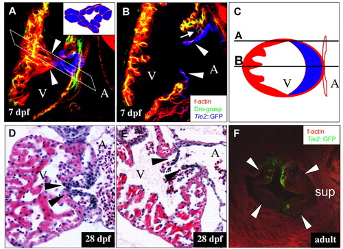
Wild-type morphogenesis of AV valve leaflets. (A,B) Confocal images of the AV canal at 7 dpf. (C) Schematic drawing of a transverse section through the ventricle at the level of a valve leaflet. (D,E) Hematoxylin and Eosin-stained sections through the AV canal at 28 dpf. (F) Adult valve leaflets. (A,B) Sections of a Tg(Tie2:EGFP)s849 (pseudo-colored blue) heart immunostained for Dm-grasp (pseudo-colored green) and stained with rhodamine phalloidin (red). (A) Optical section through the lateral wall of the ventricle as indicated in C shows that each of the two leaflets is connected to distal ventricular myocardial trabeculae (arrowheads). White parallelogram indicates the plane schematically depicted in C. (B) Optical section through the ventricle as indicated in C. Valve leaflets consist of two layers of Tg(Tie2:EGFP)s849 positive cells (arrowheads). The myocardial wall distal to the AV canal is trabeculated in contrast to the juxtavalvular ventricular wall. The superior valve leaflet (arrow) is connected to the AV myocardium by a row of weakly Tg(Tie2:EGFP)s849-positive (i.e. endocardially derived) cuboidal cells that express Dm-grasp (arrow). (C) Lines labeled A and B represent the approximate latitude of the optical sections shown in A and B, respectively. (D) Oblique section through the AV canal shows that the valve leaflets (arrowheads) are connected laterally to the ventricular trabeculae. (E) Sagittal section through the heart shows that the valve leaflets consist of two layers of cells (arrowheads). (F) The adult zebrafish AV valve consists of four leaflets (arrowheads). A, atrium; V, ventricle; sup, superior AV valve leaflets.
|
|
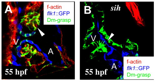
Regulation of AV endocardial development. Confocal images of the AV canal of wild-type (A) and mutant (B) hearts at 55 hpf. (A,B) Tg(flk1:EGFP)s843 (pseudo-colored blue) embryos immunostained for Dm-grasp (pseudo-colored green) and stained with rhodamine phalloidin (red). In contrast to wild type (A), in sih mutant embryos (B), AV canal endocardial cells (arrowhead) fail to express Dm-grasp and to adopt a cuboidal shape. Interestingly, the sih mutant myocardium is devoid of filamentous actin staining. A, atrium; V, ventricle.
|
|
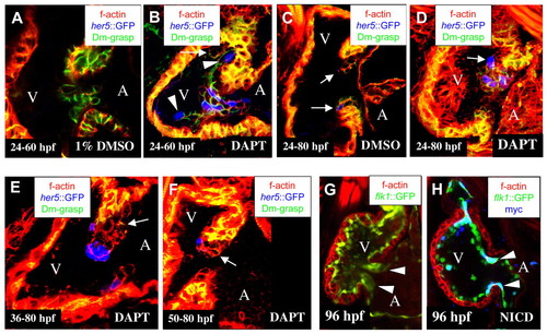
Notch signaling restricts the differentiation of cuboidal endocardium to the AV canal. Confocal images of hearts from DMSO-treated (A,C) and DAPT-treated embryos (B,D-F) at 60 (A,B) and 80 hpf (C-F), and from wild-type (G) and NotchICD-overexpressing (H) embryos at 96 hpf (G,H). (A-F) Tg(0.7her5:EGFP)ne2067 embryos (pseudo-colored blue) immunostained for Dm-grasp (pseudo-colored green) and stained with rhodamine phalloidin (red). (G,H) Tg(flk1:Gal4-UAS:EGFP)s848 (green) (G) and transheterozygous Tg(flk1:Gal4-UAS:EGFP)s848 (green); Tg(UAS:myc-Notch1a-intra)kca3 (H) embryos immunostained for MYC (blue) and stained with rhodamine phalloidin (red). (Most endocardial cells stained positive for MYC expression although the AV canal endocardial cells appear to express higher levels.) (A) In embryos treated with 1% DMSO, cuboidal, Dm-grasp positive endocardial cells were restricted to the AV canal. (B) In embryos treated with 100 µM DAPT between 24-60 hpf, the ventricular endocardium showed ectopic cuboidal, Dm-grasp-positive cells (arrow) and ectopic Tg(0.7her5:EGFP)ne2067 expression (arrowheads). (C) In embryos treated with 1% DMSO from 24-80 hpf, EC development was unaffected, and Tg(0.7her5:EGFP)ne2067 expression was restricted to single cells located at the boundary between the ventricle and AV canal (arrows). Embryos treated with 100 µM DAPT from 24-80 hpf (D) formed a hypercellular EC in the superior region of the AV canal with numerous Tg(0.7her5:EGFP)ne2067-positive cells. Embryos treated from 36-80 hpf (E) and from 50-80 hpf (F) formed a disorganized cushion similar to the control in size and cell numbers (compare with C) although with reduced Dm-grasp expression in the cuboidal endocardial cells (arrows). (G) Tg(flk1:Gal4-UAS:EGFP)s848 embryos have completed the formation of the superior and inferior AV canal ECs by 96 hpf (arrowheads). (H) NotchICD-expressing AV canal endocardial cells (arrowheads) remain squamous, resulting in a lack of AV canal ECs at 96 hpf.
EXPRESSION / LABELING:
|
|
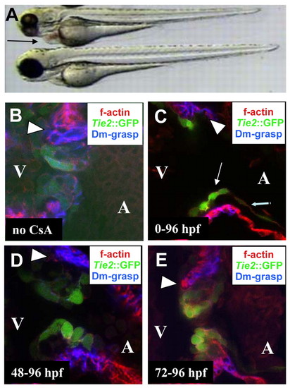
Calcineurin signaling is required for EMT and EC morphogenesis. (A) Bright-field image of an untreated embryo (bottom) and an embryo raised in 10 µg/ml CsA (top). (B-E) Confocal images of the AV canal of Tg(Tie2:EGFP)s849 (green) embryos at 96 hpf immunostained for Dm-grasp (blue) and stained with rhodamine phalloidine (red); (B) untreated embryo; (C-E) embryos treated with CsA from the one cell stage (C), 48 hpf (D) and 72 hpf (E). (A) Embryos treated with CsA from the one cell stage appeared morphologically wild type at 72 hpf, except for pericardial edema (arrow in A) owing to outflow tract stenosis (see Movie 1 in the supplementary material). (C) In embryos treated with CsA from the one-cell stage, the myocardium appeared thinner throughout the heart (compare with B, D, E and Fig. 2). AV endocardial cells upregulated Tg(Tie2:EGFP)s849 and initiated a cell shape change (thin arrow indicates AV endocardial cell, thick arrow indicates squamous atrial endocardial cell), but failed to express Dm-Grasp; no EMT occurred and ECs failed to form. (D) In embryos treated with CsA from 48 hpf, ECs appeared disorganized. (E) No effect on EMT or cushion morphogenesis was observed at 96 hpf when embryos were treated with CsA from 72 hpf onwards (compare E with B). Arrowheads indicate the superior region of the AV canal in B-E.
EXPRESSION / LABELING:
|
|
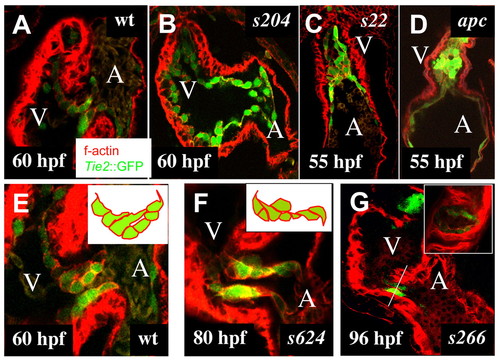
AV canal defective mutants identified in a large-scale mutagenesis screen. Wild-type (A,E) and mutant (B-D,F,G) hearts between 55 and 96 hpf. Confocal images of embryonic hearts at 60 (A,B) and 55 hpf (C,D). Confocal images of the AV canal at 60 (E), 80 (F) and 96 hpf (G). In contrast to wild-type hearts at 60 hpf (A), in s204 mutant hearts (B), the AV canal endocardial cells fail to adopt a cuboidal shape. (C) In s22 mutant hearts, the ventricular lumen contains cuboidal endocardial cells. (D) In the apc mutant heart, the ventricular lumen is filled with Tg(Tie2:EGFP)s849-positive cells with a mesenchymal morphology. (E) In wild-type embryos, only AV canal endocardial cells located next to the ventricular border form cellular extensions into the AV canal ECM and extensions are directed towards the atrial border of the AV canal. (F) In s624 mutant embryos at 80 hpf, no ECs have formed, and the AV canal endocardial cells extend cellular projections in different directions. (G) In s266 mutant embryos, no ECs have formed by 96 hpf; the AV canal is lined by a single layer of cuboidal cells. Inset shows a transverse section of the s266 mutant AV canal (compare with Fig. 2F and see Movie 2 in the supplementary material). A, atrium; V, ventricle.
|

