Fig. 1
- ID
- ZDB-FIG-050916-1
- Publication
- Beis et al., 2005 - Genetic and cellular analyses of zebrafish atrioventricular cushion and valve development
- Other Figures
- All Figure Page
- Back to All Figure Page
|
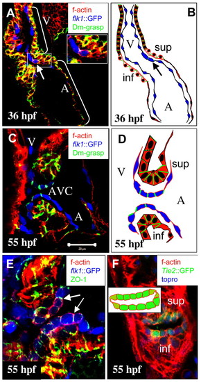
Cellular differentiation of wild-type AV canal between 36 and 55 hpf. Confocal images of the heart at 36 hpf (A) and the AV canal at 55 hpf (C,E,F). (A,C,E) Tg(flk1:EGFP)s843 (pseudo-colored blue) embryos stained with rhodamine phalloidin (red) and immunostained for Dm-grasp (pseudo-colored green) (A,C) or for ZO-1 (pseudo-colored green) (E). (F) Tg(Tie2:EGFP)s849 (green) embryo stained with topro (blue) and rhodamine phalloidin (red). (A) At 36 hpf, the heart has looped and the endocardium (in blue) is single layered and squamous. Arrow indicates one endocardial cell expressing Dm-grasp. (B) Schematic representation of the heart shown in A. (C) At 55 hpf, the AV canal endocardial cells exhibit a cuboidal shape and Dm-grasp is localized laterally. Ventricular and atrial endocardial cells appear squamous and devoid of Dm-grasp expression. Myocardial cells in the superior (sup) and inferior (inf) ECFR of the AV canal exhibit stronger expression of laterally localized Dm-grasp compared with ventricular and atrial myocardial cells. (D) Schematic representation of the AV canal shown in C. (E) ZO-1 is expressed by all myocardial and endocardial cells, including the cuboidal endocardial cells lining the AV canal. Arrows indicate ZO-1 localized in tight junctions between two neighboring cuboidal endocardial cells. (F) Transverse section. A five or six cell-wide sheet of cuboidal cells line the superior and inferior regions of the AV canal. Laterally and in this plane, two squamous cells (hinge cells; light green) connect the sheets of cuboidal cells. The inset is a schematic representation of the pattern of endocardial cell shapes across the AV canal. A, atrium; V, ventricle; AVC, atrioventricular canal; inf, inferior ECFR; sup: superior ECFR.
|
| Gene: | |
|---|---|
| Fish: | |
| Anatomical Terms: | |
| Stage Range: | Prim-25 to Long-pec |

