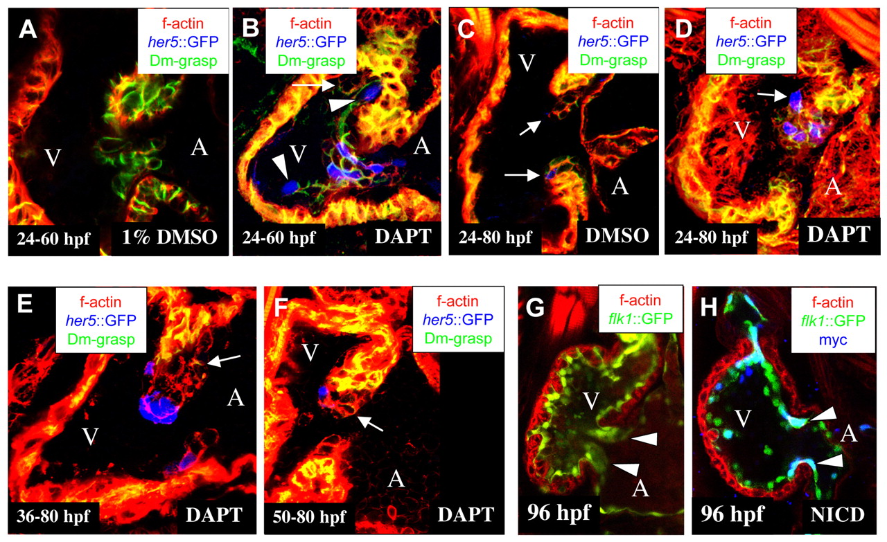
Fig. 5 Notch signaling restricts the differentiation of cuboidal endocardium to the AV canal. Confocal images of hearts from DMSO-treated (A,C) and DAPT-treated embryos (B,D-F) at 60 (A,B) and 80 hpf (C-F), and from wild-type (G) and NotchICD-overexpressing (H) embryos at 96 hpf (G,H). (A-F) Tg(0.7her5:EGFP)ne2067 embryos (pseudo-colored blue) immunostained for Dm-grasp (pseudo-colored green) and stained with rhodamine phalloidin (red). (G,H) Tg(flk1:Gal4-UAS:EGFP)s848 (green) (G) and transheterozygous Tg(flk1:Gal4-UAS:EGFP)s848 (green); Tg(UAS:myc-Notch1a-intra)kca3 (H) embryos immunostained for MYC (blue) and stained with rhodamine phalloidin (red). (Most endocardial cells stained positive for MYC expression although the AV canal endocardial cells appear to express higher levels.) (A) In embryos treated with 1% DMSO, cuboidal, Dm-grasp positive endocardial cells were restricted to the AV canal. (B) In embryos treated with 100 µM DAPT between 24-60 hpf, the ventricular endocardium showed ectopic cuboidal, Dm-grasp-positive cells (arrow) and ectopic Tg(0.7her5:EGFP)ne2067 expression (arrowheads). (C) In embryos treated with 1% DMSO from 24-80 hpf, EC development was unaffected, and Tg(0.7her5:EGFP)ne2067 expression was restricted to single cells located at the boundary between the ventricle and AV canal (arrows). Embryos treated with 100 µM DAPT from 24-80 hpf (D) formed a hypercellular EC in the superior region of the AV canal with numerous Tg(0.7her5:EGFP)ne2067-positive cells. Embryos treated from 36-80 hpf (E) and from 50-80 hpf (F) formed a disorganized cushion similar to the control in size and cell numbers (compare with C) although with reduced Dm-grasp expression in the cuboidal endocardial cells (arrows). (G) Tg(flk1:Gal4-UAS:EGFP)s848 embryos have completed the formation of the superior and inferior AV canal ECs by 96 hpf (arrowheads). (H) NotchICD-expressing AV canal endocardial cells (arrowheads) remain squamous, resulting in a lack of AV canal ECs at 96 hpf.

