Fig. 5
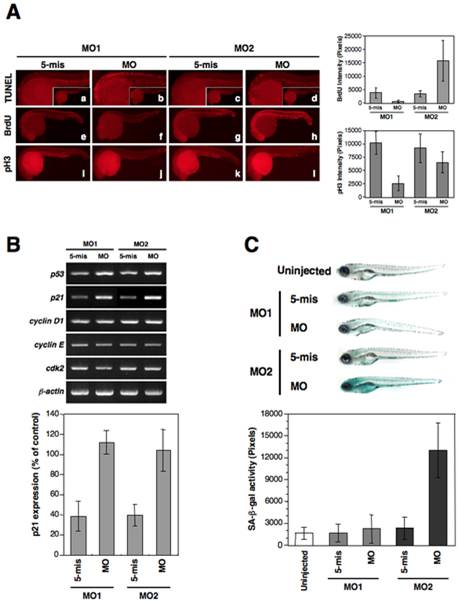
Fig. 5 The knockdown of zebrafish lamin A/C induces an aberrant cell cycle, apoptosis, and senescence.
(A) Apoptosis and cell cycle analysis in 24 hpf embryos. (a–d) Lateral view of apoptotic phenotypes in MO-injected embryos using a TUNEL assay (TUNEL-positive cells are indicated by the red fluorescence spots). zLMNA-MO1-injected embryos shows increased apoptosis in the head and trunk. (e–h) BrdU incorporation in MO-injected embryos. zLMNA-MO2-injected embryos show increased BrdU-positive cells throughout the whole body. (i–l) MO-injected embryos stained with anti-phospho histone H3 (pH 3). A zLMNA-MO1-injected embryo shows a decreased number of pH 3-positive cells. Quantifications of the BrdU and pH 3 intensities in morphants are shown in right graphs (BrdU: MO1, P<0.05 for 5-mis versus MO, MO2, P<0.05 for 5-mis versus MO; pH 3: MO1, P<0.05 for 5-mis versus MO, MO2, no significance for 5-mis versus MO). (B) Genes involved in the p53-related and cdk2 pathways were analyzed by semi-quantitative RT-PCR in MO-injected embryos at 24 hpf. Quantifications of the bands intensities for p21 are shown in the bottom graph (P<0.01 for 5-mis versus MO in MO1 and MO2). (C) SA-β-gal assay of 6 dpf MO-injected larvae in comparison with each control 5-mis MO. Qualifications of the SA-β-gal activity are shown in bottom graph. The zLMNA-MO2-injected embryos revealed strong induction of SA-β-gal activity (P<0.01 for 5-mis versus MO in MO2), whereas the zLMNA-MO1-injected embryos did not exhibit any obvious activity, compared with Cont-MO1-injected embryos.

