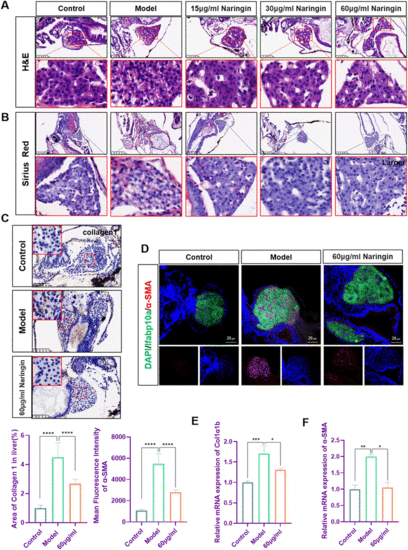
Fig. 3
|
Naringin attenuated TAA-induced liver fibrosis in zebrafish. (A) H&E staining of zebrafish larvae. Figures are magnified as ×200 (n = 8). (B) Sirius red staining of zebrafish larvae. Figures are magnified at ×100 (n = 8). (C) Immunohistochemical staining of collagen1 in paraffin sections of zebrafish larvae. Figures are magnified at ×200 (n = 8). (D) Frozen liver sections of zebrafish larvae with liver-specific eGFP expression were immunofluorescently stained with α-SMA (n = 7). (E) The qPCR analysis of Col1a1b mRNA expression in zebrafish (n = 3). (F) The qPCR analysis of α-SMA mRNA expression in zebrafish (n = 3). The mRNA expression was normalized to β-actin mRNA expression and presented as a fold change compared with the control group. ns denotes no significance, *p < 0.05, **p < 0.01, ***p < 0.001, and ****p < 0.0001. |

