
Fig. 6
- ID
- ZDB-FIG-231221-20
- Publication
- Daponte et al., 2023 - Cell differentiation and matrix organization are differentially affected during bone formation in osteogenesis imperfecta zebrafish models with different genetic defects impacting collagen type I structure
- Other Figures
- All Figure Page
- Back to All Figure Page
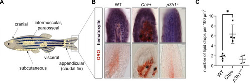
Adipocytic differentiation in the distal extremity of the caudal fin. (A) Representative images of adipocyte localization in zebrafish. Adipocytes can be found in the cranium, in the viscera, and in the subcutaneous, intermuscular and paraosseal tissues. Appendicular adipocytes were also described in the caudal fin base; vice versa, in the distal extremity of the caudal fin, few or no adipocytes should be present. Created with BioRender.com. (B) Oil Red O (ORO) and hematoxylin counterstaining of WT (n = 5), Chi/+ (n = 4) and p3h1−/− (n = 4) longitudinal caudal fin sections. Double staining on the top, ORO staining on the bottom. Scale bar: 10 µm. (C) Quantification of the number of ORO-stained lipid drops in the distal caudal fin revealed an increased adipogenesis in Chi/+ zebrafish. Each dot represents a single value: circle for WT, square for Chi/+ and triangle for p3h1−/−. * P < 0.05. |