
Fig. 2
- ID
- ZDB-FIG-231221-16
- Publication
- Daponte et al., 2023 - Cell differentiation and matrix organization are differentially affected during bone formation in osteogenesis imperfecta zebrafish models with different genetic defects impacting collagen type I structure
- Other Figures
- All Figure Page
- Back to All Figure Page
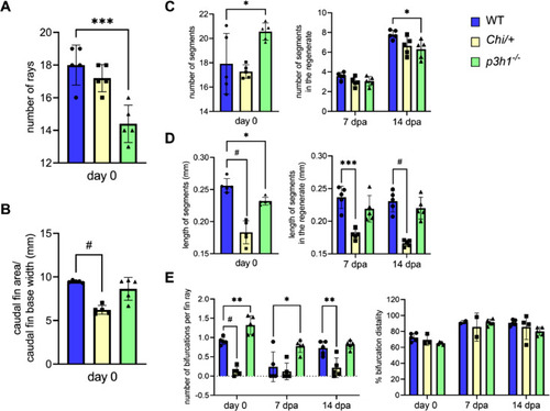
Morphometric analysis of WT, Chi/+ and p3h1−/− caudal fin before and during regeneration. Number and length of segments were measured in the total caudal fin at day 0 and only in the regenerated fin at 7 and 14 dpa. (A) A general reduction in the number of rays was detected in p3h1−/− compared to WT. (B) Caudal fin area, normalized to the fin base width, was significantly reduced in Chi/+ respect to WT. (C) At day 0 the number of segments, increased in p3h1−/− with respect to WT, was reduced in the 14 dpa regenerate, compatibly with overall reduced bone formation. (D) The medium length of segments was reduced in p3h1−/− respect to WT at day 0 and in Chi/+ respect to WT before and during regeneration. (E) p3h1−/− zebrafish presented an increased number of bifurcations at day 0 and 7 dpa, while Chi/+ rays had significantly less bifurcations at day 0 and at 14 dpa respect to WT. No differences were found in the bifurcation distality percentage, measured as the ratio between the distance to the first bifurcation and the fin ray length, between all zebrafish (n = 5 for each genotype). dpa: days post amputation. Each dot represents a single value: circle for WT, square for Chi/+ and triangle for p3h1−/−. * P < 0.05, ** P < 0.01, *** P < 0.001, # P < 0.0001. |