
Fig. 1
- ID
- ZDB-FIG-231221-15
- Publication
- Daponte et al., 2023 - Cell differentiation and matrix organization are differentially affected during bone formation in osteogenesis imperfecta zebrafish models with different genetic defects impacting collagen type I structure
- Other Figures
- All Figure Page
- Back to All Figure Page
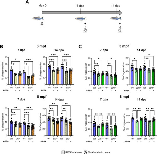
Caudal fin regeneration is reduced in Chi/+ and p3h1−/− compared to WT and is rescued by 4-PBA administration only in p3h1−/−. (A) Caudal fin was amputated and calcein vital staining was performed at 7 and 14 dpa, as shown in the scheme. (B) Caudal fin regeneration of WT and Chi/+ siblings. Chi/+ zebrafish at 3 and 8 mpf exhibited a significantly reduced caudal fin regeneration compared to WT both at 7 and 14 dpa. 4-PBA treatments did not improve either REG or EMA between controls and treated zebrafish at all ages and time points analyzed (3 mpf: WT n = 6, Chi/+ controls n = 5, treated Chi/+ n = 6; 8 mpf: WT n = 10, Chi/+ controls n = 10, treated Chi/+ n = 8). (C) Caudal fin regeneration of WT and p3h1−/− siblings. p3h1−/− mutants at 3 and 8 mpf showed a significantly reduced caudal fin regeneration compared to WT. 4-PBA administration significantly increased REG and EMA parameters in 8 mpf treated respect to controls, reaching WT value (3 mpf: WT n = 10, p3h1−/− controls n ≥ 9, treated p3h1−/− n = 10; 8 mpf: WT n = 9, p3h1−/− controls n ≥ 7, treated p3h1−/− n = 10). dpa: days post amputation; mpf: months post fertilization; REG: Regenerated Area; EMA: Estimated Mineralized Area. Each dot represents a single value: circle for WT, full square for Chi/+ controls, empty square for treated Chi/+, full triangle for p3h1−/− controls and empty triangle for treated p3h1−/−. * P < 0.05, ** P < 0.01, *** P < 0.001, # P < 0.0001. |