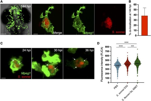
Shigella can establish persistent infection of macrophages in vivo. A, Head region and individual macrophage detail from a representative S. sonnei–infected zebrafish larva at 144 hpi. Tg(mpeg1::Gal4-FF)gl25/Tg(UAS::LIFEACT-GFP)mu271 larvae (Mpeg1, with macrophages in green) were injected at 3 dpf in the hindbrain ventricle with 1000 CFU of mCherry-labeled S. sonnei 53G (red). An infected macrophage harboring persistent bacteria is magnified. Scale bars: 100 μm, left; 10 μm, right and inset. B, At the persistent infection stage, 40% of bacterial fluorescence colocalizes with mpeg1+ macrophages. C, Individual Tg(mpeg1::Gal4-FF)gl25/Tg(UAS::LIFEACT-GFP)mu271 macrophage (Mpeg1, green) harboring mCherry–S. sonnei (red, indicated by arrows) followed for 12 hours (from 24 to 36 hpi). At 2 dpf, 1000 CFU of bacteria were delivered systemically in larvae. Scale bar: 10 μm. D, As compared with S. flexneri M90T, S. sonnei 53G induces a reduced level of caspase 1 activation in vivo in the zebrafish model. At 3 dpf, 10000 CFU of bacteria were delivered systemically in larvae. Data: mean ± SEM. Statistics: 1-way analysis of variance with Sidak correction. ***P < .001. ****P < 0.0001. CFU, colony-forming unit; dpf, days postfertilization; hpi, hours postinfection; PBS, phosphate-buffered saline.
|